 ムービー
ムービー コントローラー


+ データを開く

- 基本情報
基本情報
| 登録情報 | データベース: PDB / ID: 7y1a | ||||||
|---|---|---|---|---|---|---|---|
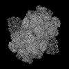
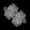
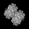
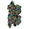
| タイトル | Lateral hexamer | ||||||
 要素 要素 |
| ||||||
 キーワード キーワード | PHOTOSYNTHESIS / Lateral hexamer / PBS | ||||||
| 機能・相同性 |  機能・相同性情報 機能・相同性情報 | ||||||
| 生物種 |  Porphyridium purpureum (真核生物) Porphyridium purpureum (真核生物) | ||||||
| 手法 | 電子顕微鏡法 / 単粒子再構成法 / クライオ電子顕微鏡法 / 解像度: 6.3 Å | ||||||
 データ登録者 データ登録者 | You, X. / Zhang, X. / Cheng, J. / Xiao, Y.N. / Sun, S. / Sui, S.F. | ||||||
| 資金援助 | 1件
| ||||||
 引用 引用 | ジャーナル: Nature / 年: 2023 タイトル: In situ structure of the red algal phycobilisome-PSII-PSI-LHC megacomplex. 著者: Xin You / Xing Zhang / Jing Cheng / Yanan Xiao / Jianfei Ma / Shan Sun / Xinzheng Zhang / Hong-Wei Wang / Sen-Fang Sui /  要旨: In oxygenic photosynthetic organisms, light energy is captured by antenna systems and transferred to photosystem II (PSII) and photosystem I (PSI) to drive photosynthesis. The antenna systems of red ...In oxygenic photosynthetic organisms, light energy is captured by antenna systems and transferred to photosystem II (PSII) and photosystem I (PSI) to drive photosynthesis. The antenna systems of red algae consist of soluble phycobilisomes (PBSs) and transmembrane light-harvesting complexes (LHCs). Excitation energy transfer pathways from PBS to photosystems remain unclear owing to the lack of structural information. Here we present in situ structures of PBS-PSII-PSI-LHC megacomplexes from the red alga Porphyridium purpureum at near-atomic resolution using cryogenic electron tomography and in situ single-particle analysis, providing interaction details between PBS, PSII and PSI. The structures reveal several unidentified and incomplete proteins and their roles in the assembly of the megacomplex, as well as a huge and sophisticated pigment network. This work provides a solid structural basis for unravelling the mechanisms of PBS-PSII-PSI-LHC megacomplex assembly, efficient energy transfer from PBS to the two photosystems, and regulation of energy distribution between PSII and PSI. | ||||||
| 履歴 |
|
- 構造の表示
構造の表示
| 構造ビューア | 分子: MolmilJmol/JSmol |
|---|
- ダウンロードとリンク
-ダウンロード
| PDBx/mmCIF形式 | 7y1a.cif.gz | 381.7 KB | 表示 | PDBx/mmCIF形式 |
|---|---|---|---|---|
| PDB形式 | pdb7y1a.ent.gz | 332.2 KB | 表示 | PDB形式 |
| PDBx/mmJSON形式 | 7y1a.json.gz | ツリー表示 | PDBx/mmJSON形式 | |
| その他 |  その他のダウンロード その他のダウンロード |
-検証レポート
| 文書・要旨 | 7y1a_validation.pdf.gz | 3.6 MB | 表示 | wwPDB検証レポート |
|---|---|---|---|---|
| 文書・詳細版 | 7y1a_full_validation.pdf.gz | 3.7 MB | 表示 | |
| XML形式データ | 7y1a_validation.xml.gz | 86.1 KB | 表示 | |
| CIF形式データ | 7y1a_validation.cif.gz | 103.1 KB | 表示 | |
| アーカイブディレクトリ | https://data.pdbj.org/pub/pdb/validation_reports/y1/7y1aftp://data.pdbj.org/pub/pdb/validation_reports/y1/7y1a | HTTPS FTP |
-関連構造データ


-リンク
 PDBj
PDBj- 集合体
集合体
| 登録構造単位 | 
|
|---|---|
| 1 |
|
-要素
| #1: タンパク質 | 分子量: 18584.182 Da / 分子数: 6 / 由来タイプ: 組換発現 由来: (組換発現) Porphyridium purpureum (真核生物)遺伝子: cpeB / 発現宿主: Porphyridium purpureum (真核生物) / 参照: UniProt: P11393#2: タンパク質 | 分子量: 17824.029 Da / 分子数: 6 / 由来タイプ: 組換発現 由来: (組換発現) Porphyridium purpureum (真核生物)遺伝子: peA, cpeA / 発現宿主: Porphyridium purpureum (真核生物) / 参照: UniProt: E2IH77#3: タンパク質 | | 分子量: 7422.140 Da / 分子数: 1 / 由来タイプ: 組換発現 由来: (組換発現) Porphyridium purpureum (真核生物)発現宿主: Porphyridium purpureum (真核生物)#4: タンパク質 | | 分子量: 12783.750 Da / 分子数: 1 / 由来タイプ: 組換発現 由来: (組換発現) Porphyridium purpureum (真核生物)発現宿主: Porphyridium purpureum (真核生物)#5: 化合物 | ChemComp-PEB /   分子量: 588.694 Da / 分子数: 31 / 由来タイプ: 合成 / 式: C33H40N4O6 / タイプ: SUBJECT OF INVESTIGATION 分子量: 588.694 Da / 分子数: 31 / 由来タイプ: 合成 / 式: C33H40N4O6 / タイプ: SUBJECT OF INVESTIGATION研究の焦点であるリガンドがあるか | Y | |
|---|
-実験情報
-実験
| 実験 | 手法: 電子顕微鏡法 |
|---|---|
| EM実験 | 試料の集合状態: CELL / 3次元再構成法: 単粒子再構成法 |
- 試料調製
試料調製
| 構成要素 | 名称: Lateral hexamer of PBS / タイプ: COMPLEX / Entity ID: #1-#4 / 由来: NATURAL |
|---|---|
| 由来(天然) | 生物種: Porphyridium purpureum (真核生物) |
| 緩衝液 | pH: 7 |
| 試料 | 包埋: NO / シャドウイング: NO / 染色: NO / 凍結: YES |
| 急速凍結 | 凍結剤: ETHANE |
- 電子顕微鏡撮影
| 実験機器 |  モデル: Titan Krios / 画像提供: FEI Company |
|---|---|
| 顕微鏡 | モデル: FEI TITAN KRIOS |
| 電子銃 | 電子線源:  FIELD EMISSION GUN / 加速電圧: 300 kV / 照射モード: SPOT SCAN FIELD EMISSION GUN / 加速電圧: 300 kV / 照射モード: SPOT SCAN |
| 電子レンズ | モード: BRIGHT FIELD / 最大 デフォーカス(公称値): 6000 nm / 最小 デフォーカス(公称値): 1000 nm |
| 撮影 | 電子線照射量: 35 e/Å2 / フィルム・検出器のモデル: GATAN K3 (6k x 4k) |
- 解析
解析
| CTF補正 | タイプ: PHASE FLIPPING AND AMPLITUDE CORRECTION |
|---|---|
| 3次元再構成 | 解像度: 6.3 Å / 解像度の算出法: FSC 0.143 CUT-OFF / 粒子像の数: 87000 / 対称性のタイプ: POINT |
