Movie
Movie Controller


+ Open data

- Basic information
| Entry |  | |||||||||
|---|---|---|---|---|---|---|---|---|---|---|
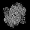
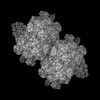
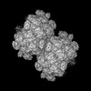
| Title | PBS of PBS-PSII-PSI-LHCs from Porphyridium purpureum. | |||||||||
 Map data Map data | ||||||||||
 Sample Sample |
| |||||||||
 Keywords Keywords | PBS / PHOTOSYNTHESIS | |||||||||
| Function / homology |  Function and homology information Function and homology informationphycobilisome / chloroplast thylakoid membrane / photosynthesis / endomembrane system / lyase activity Similarity search - Function | |||||||||
| Biological species |  Porphyridium purpureum (eukaryote) Porphyridium purpureum (eukaryote) | |||||||||
| Method | single particle reconstruction / cryo EM / Resolution: 3.3 Å | |||||||||
 Authors Authors | You X / Zhang X / Cheng J / Xiao YN / Sun S / Sui SF | |||||||||
| Funding support | 1 items
| |||||||||
 Citation Citation | Journal: Nature / Year: 2023 Title: In situ structure of the red algal phycobilisome-PSII-PSI-LHC megacomplex. Authors: Xin You / Xing Zhang / Jing Cheng / Yanan Xiao / Jianfei Ma / Shan Sun / Xinzheng Zhang / Hong-Wei Wang / Sen-Fang Sui /  Abstract: In oxygenic photosynthetic organisms, light energy is captured by antenna systems and transferred to photosystem II (PSII) and photosystem I (PSI) to drive photosynthesis. The antenna systems of red ...In oxygenic photosynthetic organisms, light energy is captured by antenna systems and transferred to photosystem II (PSII) and photosystem I (PSI) to drive photosynthesis. The antenna systems of red algae consist of soluble phycobilisomes (PBSs) and transmembrane light-harvesting complexes (LHCs). Excitation energy transfer pathways from PBS to photosystems remain unclear owing to the lack of structural information. Here we present in situ structures of PBS-PSII-PSI-LHC megacomplexes from the red alga Porphyridium purpureum at near-atomic resolution using cryogenic electron tomography and in situ single-particle analysis, providing interaction details between PBS, PSII and PSI. The structures reveal several unidentified and incomplete proteins and their roles in the assembly of the megacomplex, as well as a huge and sophisticated pigment network. This work provides a solid structural basis for unravelling the mechanisms of PBS-PSII-PSI-LHC megacomplex assembly, efficient energy transfer from PBS to the two photosystems, and regulation of energy distribution between PSII and PSI. | |||||||||
| History |
|
- Structure visualization
| Supplemental images |
|---|
- Downloads & links
-EMDB archive
| Map data | emd_33605.map.gz | 203.7 MB | EMDB map data format | |
|---|---|---|---|---|
| Header (meta data) | emd-33605-v30.xmlemd-33605.xml | 58.3 KB 58.3 KB | Display Display | EMDB header |
| Images |  emd_33605.png emd_33605.png | 183.6 KB | ||
| Filedesc metadata | emd-33605.cif.gz | 13 KB | ||
| Others | emd_33605_half_map_1.map.gzemd_33605_half_map_2.map.gz | 200.5 MB 200.5 MB | ||
| Archive directory |  http://ftp.pdbj.org/pub/emdb/structures/EMD-33605ftp://ftp.pdbj.org/pub/emdb/structures/EMD-33605 http://ftp.pdbj.org/pub/emdb/structures/EMD-33605ftp://ftp.pdbj.org/pub/emdb/structures/EMD-33605 | HTTPS FTP |
-Related structure data
| Related structure data |  7y4lMC  8jnnM  7y1aC  7y5eC  7y7aC M: atomic model generated by this map C: citing same article ( |
|---|---|
| Similar structure data |

-Links
| EMDB pages | EMDB (EBI/PDBe) / EMDataResource |
|---|
-Map
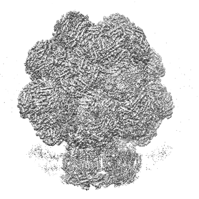
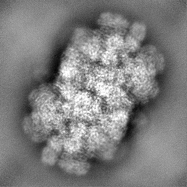
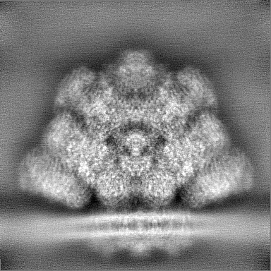
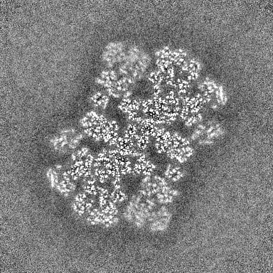
| File | Download / File: emd_33605.map.gz / Format: CCP4 / Size: 216 MB / Type: IMAGE STORED AS FLOATING POINT NUMBER (4 BYTES) | ||||||||||||||||||||||||||||||||||||
|---|---|---|---|---|---|---|---|---|---|---|---|---|---|---|---|---|---|---|---|---|---|---|---|---|---|---|---|---|---|---|---|---|---|---|---|---|---|
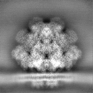
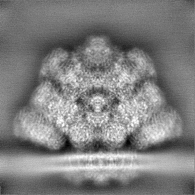
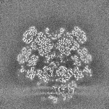
| Projections & slices | Image control
Images are generated by Spider. | ||||||||||||||||||||||||||||||||||||
| Voxel size | X=Y=Z: 1.632 Å | ||||||||||||||||||||||||||||||||||||
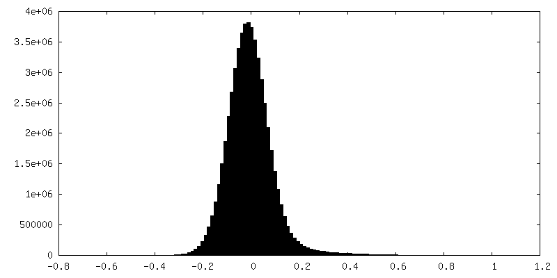
| Density |
| ||||||||||||||||||||||||||||||||||||
| Symmetry | Space group: 1 | ||||||||||||||||||||||||||||||||||||
| Details | EMDB XML:
|
 Z (Sec.)
 Y (Row.)
 X (Col.)


-Supplemental data
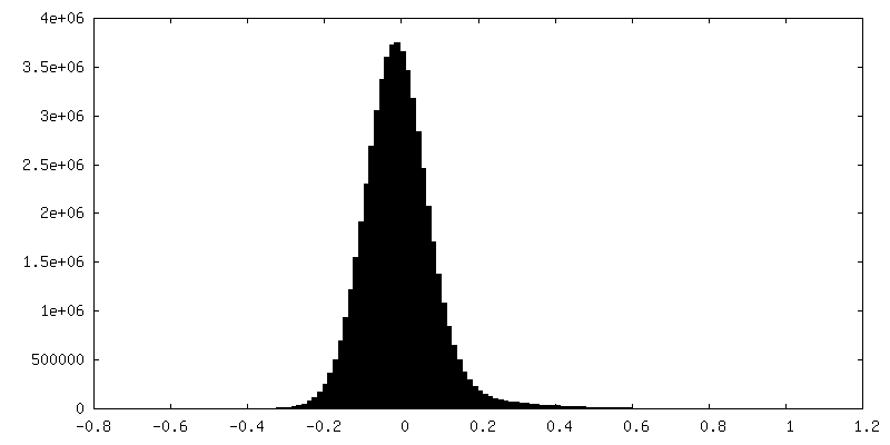
-Half map: #2
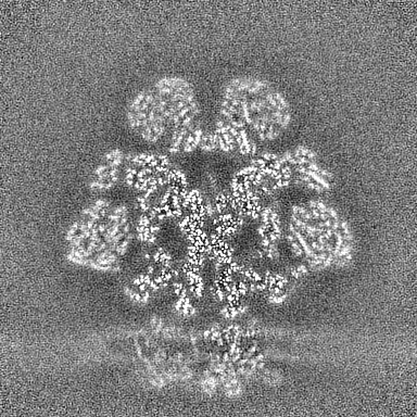
| File | emd_33605_half_map_1.map | ||||||||||||
|---|---|---|---|---|---|---|---|---|---|---|---|---|---|
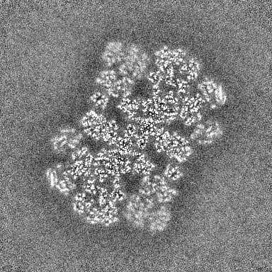
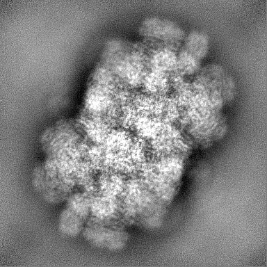
| Projections & Slices |
| ||||||||||||
| Density Histograms |

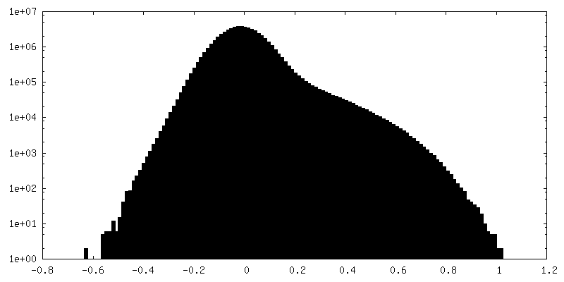
-Half map: #1
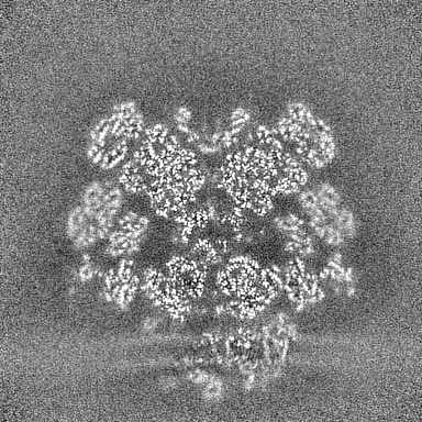
| File | emd_33605_half_map_2.map | ||||||||||||
|---|---|---|---|---|---|---|---|---|---|---|---|---|---|
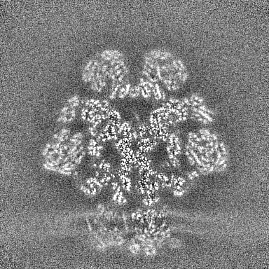
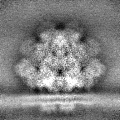
| Projections & Slices |
| ||||||||||||
| Density Histograms |
- Sample components
+Entire : In situ PBS from Porphyridium purpureum
+Supramolecule #1: In situ PBS from Porphyridium purpureum
+Macromolecule #1: Linker4
+Macromolecule #2: B-phycoerythrin beta chain
+Macromolecule #3: Phycoerythrin alpha subunit
+Macromolecule #4: CaRSPs1
+Macromolecule #5: CaRSPs2
+Macromolecule #6: R-phycoerythrin gamma chain, chloroplastic
+Macromolecule #7: Phycobilisome rod-core linker polypeptide
+Macromolecule #8: C-phycocyanin alpha subunit
+Macromolecule #9: C-phycocyanin beta subunit
+Macromolecule #10: Phycobilisome 31.8 kDa linker polypeptide, phycoerythrin-associat...
+Macromolecule #11: Phycobilisome 27.9 kDa linker polypeptide, phycoerythrin-associat...
+Macromolecule #12: R-phycoerythrin gamma chain, chloroplastic
+Macromolecule #13: R-phycoerythrin gamma chain, chloroplastic
+Macromolecule #14: R-phycoerythrin gamma chain, chloroplastic
+Macromolecule #15: Phycobilisome 31.8 kDa linker polypeptide, phycoerythrin-associat...
+Macromolecule #16: Lrc4
+Macromolecule #17: LRC5
+Macromolecule #18: FAS1 domain-containing protein
+Macromolecule #19: Allophycocyanin alpha subunit
+Macromolecule #20: Allophycocyanin beta subunit
+Macromolecule #21: Allophycocyanin gamma subunit
+Macromolecule #22: Allophycocyanin beta 18 subunit
+Macromolecule #23: Phycobilisome 7.8 kDa linker polypeptide, allophycocyanin-associa...
+Macromolecule #24: Phycobiliprotein ApcE
+Macromolecule #25: Phycobilisome 32.1 kDa linker polypeptide, phycocyanin-associated, rod
+Macromolecule #26: Phycobilisome 27.9 kDa linker polypeptide, phycoerythrin-associat...
+Macromolecule #27: Phycobilisome 31.8 kDa linker polypeptide, phycoerythrin-associat...
+Macromolecule #28: Phycobilisome 31.8 kDa linker polypeptide, phycoerythrin-associat...
+Macromolecule #29: FAS1 domain-containing protein
+Macromolecule #30: LPP2
+Macromolecule #31: PHYCOERYTHROBILIN

+Macromolecule #32: PHYCOUROBILIN
+Macromolecule #33: PHYCOCYANOBILIN

-Experimental details
-Structure determination
| Method | cryo EM |
|---|---|
 Processing Processing | single particle reconstruction |
| Aggregation state | cell |
-Sample preparation
| Buffer | pH: 7 |
|---|---|
| Vitrification | Cryogen name: ETHANE |
- Electron microscopy
| Microscope | FEI TITAN KRIOS |
|---|---|
| Image recording | Film or detector model: GATAN K3 (6k x 4k) / Average electron dose: 35.0 e/Å2 |
| Electron beam | Acceleration voltage: 300 kV / Electron source:  FIELD EMISSION GUN FIELD EMISSION GUN |
| Electron optics | Illumination mode: SPOT SCAN / Imaging mode: BRIGHT FIELD / Nominal defocus max: 6.0 µm / Nominal defocus min: 1.0 µm |
| Experimental equipment |  Model: Titan Krios / Image courtesy: FEI Company |