 Movie
Movie Controller


+ Open data

- Basic information
| Entry | Database: PDB / ID: 7xx2 | |||||||||||||||||||||||||||||||||
|---|---|---|---|---|---|---|---|---|---|---|---|---|---|---|---|---|---|---|---|---|---|---|---|---|---|---|---|---|---|---|---|---|---|---|
| Title | Cryo-EM structure of Sr35 resistosome induced by AvrSr35 R381A | |||||||||||||||||||||||||||||||||
 Components Components |
| |||||||||||||||||||||||||||||||||
 Keywords Keywords | PLANT PROTEIN / Plant immunity / NLR / resistosome | |||||||||||||||||||||||||||||||||
| Function / homology |  Function and homology information Function and homology informationplant-type hypersensitive response / innate immune response-activating signaling pathway / ADP binding / defense response to bacterium Similarity search - Function | |||||||||||||||||||||||||||||||||
| Biological species |   Puccinia graminis f. sp. tritici (fungus) Puccinia graminis f. sp. tritici (fungus) | |||||||||||||||||||||||||||||||||
| Method | ELECTRON MICROSCOPY / single particle reconstruction / cryo EM / Resolution: 3.6 Å | |||||||||||||||||||||||||||||||||
 Authors Authors | Ouyang, S.Y. / Zhao, Y.B. / Li, Z.K. / Liu, M.X. | |||||||||||||||||||||||||||||||||
| Funding support |  China, 1items China, 1items
| |||||||||||||||||||||||||||||||||
 Citation Citation | Journal: Sci Adv / Year: 2022 Title: Pathogen effector AvrSr35 triggers Sr35 resistosome assembly via a direct recognition mechanism. Authors: Yan-Bo Zhao / Meng-Xi Liu / Tao-Tao Chen / Xiaomin Ma / Ze-Kai Li / Zichao Zheng / Si-Ru Zheng / Lifei Chen / You-Zhi Li / Li-Rui Tang / Qi Chen / Peiyi Wang / Songying Ouyang / Abstract: Nucleotide-binding, leucine-rich repeat receptors (NLRs) perceive pathogen effectors to trigger plant immunity. The direct recognition mechanism of pathogen effectors by coiled-coil NLRs (CNLs) ...Nucleotide-binding, leucine-rich repeat receptors (NLRs) perceive pathogen effectors to trigger plant immunity. The direct recognition mechanism of pathogen effectors by coiled-coil NLRs (CNLs) remains unclear. We demonstrate that the CNL Sr35 directly recognizes the pathogen effector AvrSr35 from f. sp and report a cryo-electron microscopy structure of Sr35 resistosome and a crystal structure of AvrSr35. We show that AvrSr35 forms homodimers that are disassociated into monomers upon direct recognition by the leucine-rich repeat domain of Sr35, which induces Sr35 resistosome assembly and the subsequent immune response. The first 20 amino-terminal residues of Sr35 are indispensable for immune signaling but not for plasma membrane association. Our findings reveal the direct recognition and activation mechanism of a plant CNL and provide insights into biochemical function of Sr35 resistosome. | |||||||||||||||||||||||||||||||||
| History |
|
- Structure visualization
| Structure viewer | Molecule: MolmilJmol/JSmol |
|---|
- Downloads & links
-Download
| PDBx/mmCIF format | 7xx2.cif.gz | 803.5 KB | Display | PDBx/mmCIF format |
|---|---|---|---|---|
| PDB format | pdb7xx2.ent.gz | 650.8 KB | Display | PDB format |
| PDBx/mmJSON format | 7xx2.json.gz | Tree view | PDBx/mmJSON format | |
| Others |  Other downloads Other downloads |
-Validation report
| Arichive directory | https://data.pdbj.org/pub/pdb/validation_reports/xx/7xx2ftp://data.pdbj.org/pub/pdb/validation_reports/xx/7xx2 | HTTPS FTP |
|---|
-Related structure data
| Related structure data |  33498MC 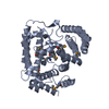 7xdsC  7xe0C  7xvgC M: map data used to model this data C: citing same article ( |
|---|---|
| Similar structure data |


-Links
 PDBj
PDBj
- Assembly
| Deposited unit | 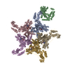
|
|---|---|
| 1 |
|
-Components
| #1: Protein | Mass: 105392.203 Da / Num. of mol.: 5 Source method: isolated from a genetically manipulated source Source: (gene. exp.) Production host: Insect expression vector pBlueBacmsGCB1 (others) References: UniProt: S5ABD6 #2: Protein | | Mass: 66019.477 Da / Num. of mol.: 1 / Mutation: R381A Source method: isolated from a genetically manipulated source Source: (gene. exp.) Puccinia graminis f. sp. tritici (fungus)Gene: PGT21_019944, PGTUg99_030428 Production host:  References: UniProt: A0A5B0N367 #3: Chemical | ChemComp-ATP /   Mass: 507.181 Da / Num. of mol.: 5 / Source method: obtained synthetically / Formula: C10H16N5O13P3 / Feature type: SUBJECT OF INVESTIGATION / Comment: ATP, energy-carrying molecule*YM Mass: 507.181 Da / Num. of mol.: 5 / Source method: obtained synthetically / Formula: C10H16N5O13P3 / Feature type: SUBJECT OF INVESTIGATION / Comment: ATP, energy-carrying molecule*YMHas ligand of interest | Y | Has protein modification | N | |
|---|
-Experimental details
-Experiment
| Experiment | Method: ELECTRON MICROSCOPY |
|---|---|
| EM experiment | Aggregation state: PARTICLE / 3D reconstruction method: single particle reconstruction |
- Sample preparation
| Component |
| ||||||||||||||||||||||||
|---|---|---|---|---|---|---|---|---|---|---|---|---|---|---|---|---|---|---|---|---|---|---|---|---|---|
| Source (natural) |
| ||||||||||||||||||||||||
| Source (recombinant) |
| ||||||||||||||||||||||||
| Buffer solution | pH: 7.5 | ||||||||||||||||||||||||
| Specimen | Embedding applied: NO / Shadowing applied: NO / Staining applied: NO / Vitrification applied: YES | ||||||||||||||||||||||||
| Vitrification | Cryogen name: ETHANE |
- Electron microscopy imaging
| Microscopy | Model: FEI TITAN |
|---|---|
| Electron gun | Electron source:  FIELD EMISSION GUN / Accelerating voltage: 300 kV / Illumination mode: FLOOD BEAM FIELD EMISSION GUN / Accelerating voltage: 300 kV / Illumination mode: FLOOD BEAM |
| Electron lens | Mode: BRIGHT FIELD / Nominal defocus max: 1600 nm / Nominal defocus min: 1000 nm |
| Image recording | Electron dose: 50 e/Å2 / Film or detector model: GATAN K3 BIOQUANTUM (6k x 4k) |
- Processing
| Software | Name: PHENIX / Version: 1.19.2_4158: / Classification: refinement | ||||||||||||||||||||||||
|---|---|---|---|---|---|---|---|---|---|---|---|---|---|---|---|---|---|---|---|---|---|---|---|---|---|
| EM software | Name: PHENIX / Category: model refinement | ||||||||||||||||||||||||
| CTF correction | Type: PHASE FLIPPING AND AMPLITUDE CORRECTION | ||||||||||||||||||||||||
| 3D reconstruction | Resolution: 3.6 Å / Resolution method: FSC 0.143 CUT-OFF / Num. of particles: 30530 / Symmetry type: POINT | ||||||||||||||||||||||||
| Refine LS restraints |
|
