 Movie
Movie Controller


+ Open data

- Basic information
| Entry | Database: PDB / ID: 7xke | |||||||||||||||||||||||||||||||||||||||||||||
|---|---|---|---|---|---|---|---|---|---|---|---|---|---|---|---|---|---|---|---|---|---|---|---|---|---|---|---|---|---|---|---|---|---|---|---|---|---|---|---|---|---|---|---|---|---|---|
| Title | Cryo-EM structure of DHEA-ADGRG2-FL-Gs complex | |||||||||||||||||||||||||||||||||||||||||||||
 Components Components |
| |||||||||||||||||||||||||||||||||||||||||||||
 Keywords Keywords | MEMBRANE PROTEIN / ADGRG2 / GPCR | |||||||||||||||||||||||||||||||||||||||||||||
| Function / homology |  Function and homology information Function and homology informationspermatid development / G protein-coupled receptor activity / Olfactory Signaling Pathway / Activation of the phototransduction cascade / G protein-coupled acetylcholine receptor signaling pathway / G beta:gamma signalling through PLC beta / Presynaptic function of Kainate receptors / Thromboxane signalling through TP receptor / Activation of G protein gated Potassium channels / Inhibition of voltage gated Ca2+ channels via Gbeta/gamma subunits ...spermatid development / G protein-coupled receptor activity / Olfactory Signaling Pathway / Activation of the phototransduction cascade / G protein-coupled acetylcholine receptor signaling pathway / G beta:gamma signalling through PLC beta / Presynaptic function of Kainate receptors / Thromboxane signalling through TP receptor / Activation of G protein gated Potassium channels / Inhibition of voltage gated Ca2+ channels via Gbeta/gamma subunits / G-protein activation / Glucagon signaling in metabolic regulation / Prostacyclin signalling through prostacyclin receptor / G beta:gamma signalling through CDC42 / Synthesis, secretion, and inactivation of Glucagon-like Peptide-1 (GLP-1) / G beta:gamma signalling through BTK / photoreceptor disc membrane / ADP signalling through P2Y purinoceptor 12 / Glucagon-type ligand receptors / Sensory perception of sweet, bitter, and umami (glutamate) taste / Adrenaline,noradrenaline inhibits insulin secretion / Vasopressin regulates renal water homeostasis via Aquaporins / Glucagon-like Peptide-1 (GLP1) regulates insulin secretion / G alpha (z) signalling events / cellular response to catecholamine stimulus / ADP signalling through P2Y purinoceptor 1 / ADORA2B mediated anti-inflammatory cytokines production / G beta:gamma signalling through PI3Kgamma / adenylate cyclase-activating dopamine receptor signaling pathway / Cooperation of PDCL (PhLP1) and TRiC/CCT in G-protein beta folding / GPER1 signaling / cellular response to prostaglandin E stimulus / heterotrimeric G-protein complex / G alpha (12/13) signalling events / G-protein beta-subunit binding / Inactivation, recovery and regulation of the phototransduction cascade / extracellular vesicle / sensory perception of taste / Thrombin signalling through proteinase activated receptors (PARs) / adenylate cyclase-activating G protein-coupled receptor signaling pathway / signaling receptor complex adaptor activity / retina development in camera-type eye / GTPase binding / fibroblast proliferation / Ca2+ pathway / High laminar flow shear stress activates signaling by PIEZO1 and PECAM1:CDH5:KDR in endothelial cells / G alpha (i) signalling events / G alpha (s) signalling events / phospholipase C-activating G protein-coupled receptor signaling pathway / G alpha (q) signalling events / Ras protein signal transduction / cell surface receptor signaling pathway / Extra-nuclear estrogen signaling / cell population proliferation / apical plasma membrane / G protein-coupled receptor signaling pathway / lysosomal membrane / GTPase activity / synapse / protein-containing complex binding / signal transduction / extracellular exosome / membrane / plasma membrane / cytoplasm / cytosol Similarity search - Function | |||||||||||||||||||||||||||||||||||||||||||||
| Biological species |  Homo sapiens (human) Homo sapiens (human) | |||||||||||||||||||||||||||||||||||||||||||||
| Method | ELECTRON MICROSCOPY / single particle reconstruction / cryo EM / Resolution: 2.9 Å | |||||||||||||||||||||||||||||||||||||||||||||
 Authors Authors | Guo, S.C. / Xiao, P. / Lin, H. / Sun, J.P. / Yu, X. | |||||||||||||||||||||||||||||||||||||||||||||
| Funding support |  China, 4items China, 4items
| |||||||||||||||||||||||||||||||||||||||||||||
 Citation Citation | Journal: Nat Chem Biol / Year: 2022 Title: Structures of the ADGRG2-G complex in apo and ligand-bound forms. Authors: Hui Lin / Peng Xiao / Rui-Qian Bu / Shengchao Guo / Zhao Yang / Daopeng Yuan / Zhong-Liang Zhu / Chuan-Xin Zhang / Qing-Tao He / Chao Zhang / Yu-Qi Ping / Ru-Jia Zhao / Chuan-Shun Ma / Chang- ...Authors: Hui Lin / Peng Xiao / Rui-Qian Bu / Shengchao Guo / Zhao Yang / Daopeng Yuan / Zhong-Liang Zhu / Chuan-Xin Zhang / Qing-Tao He / Chao Zhang / Yu-Qi Ping / Ru-Jia Zhao / Chuan-Shun Ma / Chang-Hao Liu / Xiao-Ning Zhang / Dan Jiang / Shaohui Huang / Yue-Tong Xi / Dao-Lai Zhang / Chen-Yang Xue / Bai-Sheng Yang / Jian-Yuan Li / Hao-Cheng Lin / Xu-Hui Zeng / Han Zhao / Wen-Ming Xu / Fan Yi / Zhongmin Liu / Jin-Peng Sun / Xiao Yu / Abstract: Adhesion G protein-coupled receptors are elusive in terms of their structural information and ligands. Here, we solved the cryogenic-electron microscopy (cryo-EM) structure of apo-ADGRG2, an ...Adhesion G protein-coupled receptors are elusive in terms of their structural information and ligands. Here, we solved the cryogenic-electron microscopy (cryo-EM) structure of apo-ADGRG2, an essential membrane receptor for maintaining male fertility, in complex with a G trimer. Whereas the formations of two kinks were determinants of the active state, identification of a potential ligand-binding pocket in ADGRG2 facilitated the screening and identification of dehydroepiandrosterone (DHEA), dehydroepiandrosterone sulfate and deoxycorticosterone as potential ligands of ADGRG2. The cryo-EM structures of DHEA-ADGRG2-G provided interaction details for DHEA within the seven transmembrane domains of ADGRG2. Collectively, our data provide a structural basis for the activation and signaling of ADGRG2, as well as characterization of steroid hormones as ADGRG2 ligands, which might be used as useful tools for further functional studies of the orphan ADGRG2. | |||||||||||||||||||||||||||||||||||||||||||||
| History |
|
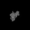
- Structure visualization
| Structure viewer | Molecule: MolmilJmol/JSmol |
|---|
- Downloads & links
-Download
| PDBx/mmCIF format | 7xke.cif.gz | 239.5 KB | Display | PDBx/mmCIF format |
|---|---|---|---|---|
| PDB format | pdb7xke.ent.gz | 170.2 KB | Display | PDB format |
| PDBx/mmJSON format | 7xke.json.gz | Tree view | PDBx/mmJSON format | |
| Others |  Other downloads Other downloads |
-Validation report
| Arichive directory | https://data.pdbj.org/pub/pdb/validation_reports/xk/7xkeftp://data.pdbj.org/pub/pdb/validation_reports/xk/7xke | HTTPS FTP |
|---|
-Related structure data
| Related structure data | 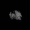 33249MC  7xkdC  7xkfC M: map data used to model this data C: citing same article ( |
|---|---|
| Similar structure data |

-Links
 PDBj
PDBj


- Assembly
| Deposited unit | 
|
|---|---|
| 1 |
|
-Components
-Protein , 2 types, 2 molecules AR
| #1: Protein | Mass: 41879.465 Da / Num. of mol.: 1 Source method: isolated from a genetically manipulated source Source: (gene. exp.) Homo sapiens (human) / Production host:  Baculovirus expression vector pFastBac1-HM Baculovirus expression vector pFastBac1-HM |
|---|---|
| #5: Protein | Mass: 98934.797 Da / Num. of mol.: 1 / Mutation: H597A, T599A Source method: isolated from a genetically manipulated source Details: C-terminal(892-916) is due to the replacement of GPR120 tail. Source: (gene. exp.) Baculovirus expression vector pFastBac1-HM / References: UniProt: Q8CJ12 |
-Guanine nucleotide-binding protein ... , 2 types, 2 molecules BG
| #2: Protein | Mass: 39489.160 Da / Num. of mol.: 1 Source method: isolated from a genetically manipulated source Source: (gene. exp.) Homo sapiens (human) / Gene: GNB1 / Production host: Baculovirus expression vector pFastBac1-HM / References: UniProt: P62873 |
|---|---|
| #3: Protein | Mass: 6375.332 Da / Num. of mol.: 1 Source method: isolated from a genetically manipulated source Source: (gene. exp.) Homo sapiens (human) / Gene: GNG2 / Production host: Baculovirus expression vector pFastBac1-HM / References: UniProt: P59768 |
-Antibody , 1 types, 1 molecules N
| #4: Antibody | Mass: 13885.439 Da / Num. of mol.: 1 Source method: isolated from a genetically manipulated source Source: (gene. exp.)  |
|---|
-Non-polymers , 2 types, 2 molecules 

| #6: Chemical | ChemComp-AND /  Mass: 288.424 Da / Num. of mol.: 1 / Source method: obtained synthetically / Formula: C19H28O2 / Feature type: SUBJECT OF INVESTIGATION / Comment: hormone*YM Mass: 288.424 Da / Num. of mol.: 1 / Source method: obtained synthetically / Formula: C19H28O2 / Feature type: SUBJECT OF INVESTIGATION / Comment: hormone*YM |
|---|---|
| #7: Water | ChemComp-HOH / Mass: 18.015 Da / Num. of mol.: 1 / Source method: isolated from a natural source / Formula: H2O |
-Details
| Has ligand of interest | Y |
|---|---|
| Has protein modification | Y |
-Experimental details
-Experiment
| Experiment | Method: ELECTRON MICROSCOPY |
|---|---|
| EM experiment | Aggregation state: PARTICLE / 3D reconstruction method: single particle reconstruction |
- Sample preparation
| Component | Name: Adhesion G-protein coupled receptor G2 / Type: COMPLEX / Entity ID: #1-#5 / Source: RECOMBINANT |
|---|---|
| Molecular weight | Value: 0.22 MDa / Experimental value: YES |
| Source (natural) | Organism: |
| Source (recombinant) | Organism: Baculovirus expression vector pFastBac1-HM |
| Buffer solution | pH: 7.5 |
| Specimen | Embedding applied: NO / Shadowing applied: NO / Staining applied: NO / Vitrification applied: YES |
| Vitrification | Cryogen name: ETHANE |
- Electron microscopy imaging
| Experimental equipment |  Model: Titan Krios / Image courtesy: FEI Company |
|---|---|
| Microscopy | Model: FEI TITAN KRIOS |
| Electron gun | Electron source:  FIELD EMISSION GUN / Accelerating voltage: 300 kV / Illumination mode: SPOT SCAN FIELD EMISSION GUN / Accelerating voltage: 300 kV / Illumination mode: SPOT SCAN |
| Electron lens | Mode: BRIGHT FIELD / Nominal defocus max: 2000 nm / Nominal defocus min: 1000 nm |
| Image recording | Electron dose: 58 e/Å2 / Film or detector model: GATAN K2 IS (4k x 4k) |
- Processing
| Software | Name: PHENIX / Version: 1.19.2_4158: / Classification: refinement | ||||||||||||||||||||||||
|---|---|---|---|---|---|---|---|---|---|---|---|---|---|---|---|---|---|---|---|---|---|---|---|---|---|
| EM software | Name: PHENIX / Category: model refinement | ||||||||||||||||||||||||
| CTF correction | Type: PHASE FLIPPING AND AMPLITUDE CORRECTION | ||||||||||||||||||||||||
| 3D reconstruction | Resolution: 2.9 Å / Resolution method: FSC 0.143 CUT-OFF / Num. of particles: 238405 / Symmetry type: POINT | ||||||||||||||||||||||||
| Refine LS restraints |
|
