National Natural Science Foundation of China (NSFC)
92057121
China
National Natural Science Foundation of China (NSFC)
32130055
China
National Science Foundation (NSF, China)
81825022
China
National Natural Science Foundation of China (NSFC)
ZR2021ZD18
China
Citation
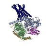
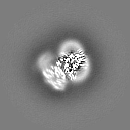
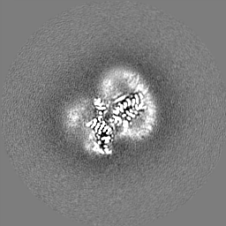
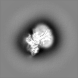
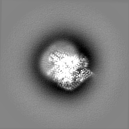
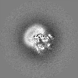
Journal: Nat Chem Biol / Year: 2022 Title: Structures of the ADGRG2-G complex in apo and ligand-bound forms. Authors: Hui Lin / Peng Xiao / Rui-Qian Bu / Shengchao Guo / Zhao Yang / Daopeng Yuan / Zhong-Liang Zhu / Chuan-Xin Zhang / Qing-Tao He / Chao Zhang / Yu-Qi Ping / Ru-Jia Zhao / Chuan-Shun Ma / Chang- ...Authors: Hui Lin / Peng Xiao / Rui-Qian Bu / Shengchao Guo / Zhao Yang / Daopeng Yuan / Zhong-Liang Zhu / Chuan-Xin Zhang / Qing-Tao He / Chao Zhang / Yu-Qi Ping / Ru-Jia Zhao / Chuan-Shun Ma / Chang-Hao Liu / Xiao-Ning Zhang / Dan Jiang / Shaohui Huang / Yue-Tong Xi / Dao-Lai Zhang / Chen-Yang Xue / Bai-Sheng Yang / Jian-Yuan Li / Hao-Cheng Lin / Xu-Hui Zeng / Han Zhao / Wen-Ming Xu / Fan Yi / Zhongmin Liu / Jin-Peng Sun / Xiao Yu / Abstract: Adhesion G protein-coupled receptors are elusive in terms of their structural information and ligands. Here, we solved the cryogenic-electron microscopy (cryo-EM) structure of apo-ADGRG2, an ...Adhesion G protein-coupled receptors are elusive in terms of their structural information and ligands. Here, we solved the cryogenic-electron microscopy (cryo-EM) structure of apo-ADGRG2, an essential membrane receptor for maintaining male fertility, in complex with a G trimer. Whereas the formations of two kinks were determinants of the active state, identification of a potential ligand-binding pocket in ADGRG2 facilitated the screening and identification of dehydroepiandrosterone (DHEA), dehydroepiandrosterone sulfate and deoxycorticosterone as potential ligands of ADGRG2. The cryo-EM structures of DHEA-ADGRG2-G provided interaction details for DHEA within the seven transmembrane domains of ADGRG2. Collectively, our data provide a structural basis for the activation and signaling of ADGRG2, as well as characterization of steroid hormones as ADGRG2 ligands, which might be used as useful tools for further functional studies of the orphan ADGRG2.
In the structure databanks used in Yorodumi, some data are registered as the other names, "COVID-19 virus" and "2019-nCoV". Here are the details of the virus and the list of structure data.
Jan 31, 2019. EMDB accession codes are about to change! (news from PDBe EMDB page)
EMDB accession codes are about to change! (news from PDBe EMDB page)
The allocation of 4 digits for EMDB accession codes will soon come to an end. Whilst these codes will remain in use, new EMDB accession codes will include an additional digit and will expand incrementally as the available range of codes is exhausted. The current 4-digit format prefixed with “EMD-” (i.e. EMD-XXXX) will advance to a 5-digit format (i.e. EMD-XXXXX), and so on. It is currently estimated that the 4-digit codes will be depleted around Spring 2019, at which point the 5-digit format will come into force.
The EM Navigator/Yorodumi systems omit the EMD- prefix.
Related info.:Q: What is EMD? / ID/Accession-code notation in Yorodumi/EM Navigator
Yorodumi is a browser for structure data from EMDB, PDB, SASBDB, etc.
This page is also the successor to EM Navigator detail page, and also detail information page/front-end page for Omokage search.
The word "yorodu" (or yorozu) is an old Japanese word meaning "ten thousand". "mi" (miru) is to see.
Related info.:EMDB / PDB / SASBDB / Comparison of 3 databanks / Yorodumi Search / Aug 31, 2016. New EM Navigator & Yorodumi / Yorodumi Papers / Jmol/JSmol / Function and homology information / Changes in new EM Navigator and Yorodumi
 Movie
Movie Controller


 Open data
 Basic information
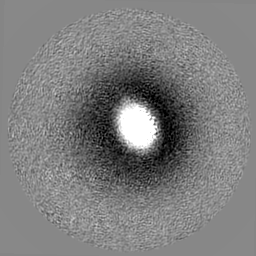
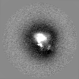
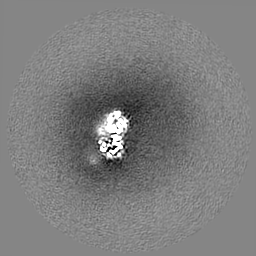
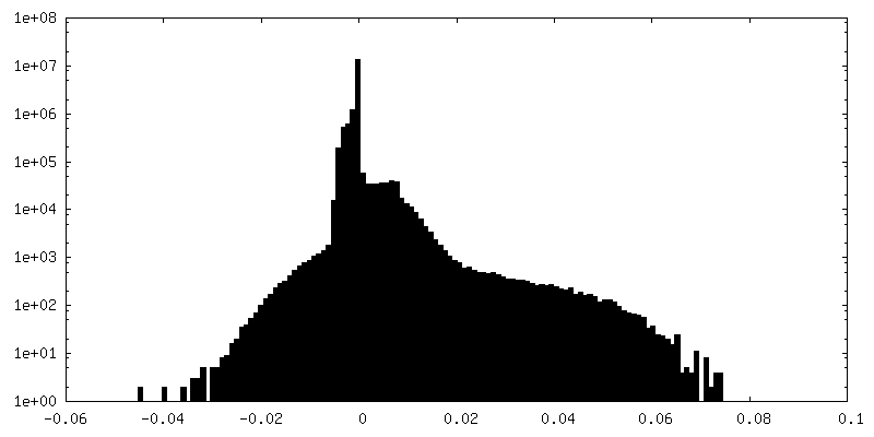
 Map data
 Sample
 Keywords
 Function and homology information
 Homo sapiens (human) /
 Authors
 China, 4 items
  Citation
 Structure visualization
 Downloads & links
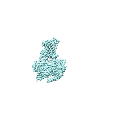 emd_33250.png
 http://ftp.pdbj.org/pub/emdb/structures/EMD-33250


 Z (Sec.)
 Y (Row.)
 X (Col.)


 Sample components
 Baculovirus expression vector pFastBac1-HM

 Processing
 Electron microscopy
 FIELD EMISSION GUN

