 Movie
Movie Controller

[English] 日本語
 Yorodumi
- PDB-7xhl: Complex structure of a Glucose 6-Phosphate Dehydrogenase from Zym... -
+ Open data

- Basic information
| Entry | Database: PDB / ID: 7xhl | ||||||
|---|---|---|---|---|---|---|---|
| Title | Complex structure of a Glucose 6-Phosphate Dehydrogenase from Zymomonas mobilis | ||||||
 Components Components | Glucose 6-Phosphate Dehydrogenase | ||||||
 Keywords Keywords | OXIDOREDUCTASE / Nicotinamide cofactor / Complex | ||||||
| Function / homology | Dihydrodipicolinate Reductase; domain 2 / Dihydrodipicolinate Reductase; domain 2 / NAD(P)-binding Rossmann-like Domain / Rossmann fold / 2-Layer Sandwich / 3-Layer(aba) Sandwich / Alpha Beta / BETA-NICOTINAMIDE RIBOSE MONOPHOSPHATE Function and homology information Function and homology information | ||||||
| Biological species |  Zymomonas mobilis (bacteria) Zymomonas mobilis (bacteria) | ||||||
| Method |  X-RAY DIFFRACTION / SYNCHROTRON / MOLECULAR REPLACEMENT / Resolution: 3.25 Å X-RAY DIFFRACTION / SYNCHROTRON / MOLECULAR REPLACEMENT / Resolution: 3.25 Å | ||||||
 Authors Authors | Meng, D.D. / Liu, M.X. / Liu, W.D. / You, C. | ||||||
| Funding support |  China, 1items China, 1items
| ||||||
 Citation Citation | Journal: Acs Catalysis / Year: 2023 Title: Coenzyme Engineering of Glucose-6-phosphate Dehydrogenase on a Nicotinamide-Based Biomimic and Its Application as a Glucose Biosensor Authors: Meng, D.D. / Liu, M.X. / Su, H. / Song, H.Y. / Chen, L.J. / Li, Q.Z. / Liu, Y.N. / Zhu, Z.G. / Liu, W.D. / Sheng, X. / You, C. / Zhang, Y.H. | ||||||
| History |
|
- Structure visualization
| Structure viewer | Molecule: MolmilJmol/JSmol |
|---|
- Downloads & links
-Download
| PDBx/mmCIF format | 7xhl.cif.gz | 701 KB | Display | PDBx/mmCIF format |
|---|---|---|---|---|
| PDB format | pdb7xhl.ent.gz | 574.2 KB | Display | PDB format |
| PDBx/mmJSON format | 7xhl.json.gz | Tree view | PDBx/mmJSON format | |
| Others |  Other downloads Other downloads |
-Validation report
| Arichive directory | https://data.pdbj.org/pub/pdb/validation_reports/xh/7xhlftp://data.pdbj.org/pub/pdb/validation_reports/xh/7xhl | HTTPS FTP |
|---|
-Related structure data
| Related structure data | 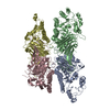 7xhpC 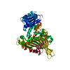 6e08S S: Starting model for refinement C: citing same article ( |
|---|---|
| Similar structure data |
-Links
 PDBj
PDBj- Assembly
| Deposited unit | 
| ||||||||
|---|---|---|---|---|---|---|---|---|---|
| 1 | 
| ||||||||
| 2 | 
| ||||||||
| Unit cell |
|
-Components
| #1: Protein | Mass: 54152.527 Da / Num. of mol.: 8 Source method: isolated from a genetically manipulated source Source: (gene. exp.) Zymomonas mobilis (bacteria) / Production host: #2: Chemical | ChemComp-NMN / 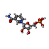  Mass: 335.227 Da / Num. of mol.: 4 / Source method: obtained synthetically / Formula: C11H16N2O8P / Feature type: SUBJECT OF INVESTIGATION Mass: 335.227 Da / Num. of mol.: 4 / Source method: obtained synthetically / Formula: C11H16N2O8P / Feature type: SUBJECT OF INVESTIGATIONHas ligand of interest | Y | |
|---|
-Experimental details
-Experiment
| Experiment | Method: X-RAY DIFFRACTION / Number of used crystals: 1 |
|---|
- Sample preparation
| Crystal | Density Matthews: 3.28 Å3/Da / Density % sol: 62.46 % |
|---|---|
| Crystal grow | Temperature: 289 K / Method: vapor diffusion, sitting drop / pH: 8.5 Details: 0.2 M di-Ammonium hydrogen Phosphate, 0.1 M Tris Hydrochloride pH 8.5, 14% w/v PEG 6000 |
-Data collection
| Diffraction | Mean temperature: 100 K / Serial crystal experiment: N | ||||||||||||||||||||||||||||||
|---|---|---|---|---|---|---|---|---|---|---|---|---|---|---|---|---|---|---|---|---|---|---|---|---|---|---|---|---|---|---|---|
| Diffraction source | Source: SYNCHROTRON / Site: SSRF / Beamline: BL18U1 / Wavelength: 0.9791 Å | ||||||||||||||||||||||||||||||
| Detector | Type: DECTRIS PILATUS3 6M / Detector: PIXEL / Date: Feb 5, 2021 | ||||||||||||||||||||||||||||||
| Radiation | Protocol: SINGLE WAVELENGTH / Monochromatic (M) / Laue (L): M / Scattering type: x-ray | ||||||||||||||||||||||||||||||
| Radiation wavelength | Wavelength: 0.9791 Å / Relative weight: 1 | ||||||||||||||||||||||||||||||
| Reflection twin | Operator: h,-k,-l / Fraction: 0.2 | ||||||||||||||||||||||||||||||
| Reflection | Resolution: 3.25→88.41 Å / Num. obs: 86216 / % possible obs: 98.9 % / Redundancy: 3.1 % / CC1/2: 0.976 / Rmerge(I) obs: 0.148 / Rpim(I) all: 0.1 / Rrim(I) all: 0.18 / Net I/σ(I): 6.4 / Num. measured all: 269015 / Scaling rejects: 80 | ||||||||||||||||||||||||||||||
| Reflection shell | Diffraction-ID: 1
|
- Processing
| Software |
| |||||||||||||||||||||||||||||||||||||||||||||||||||||||||||||||||||||||||||||||||||||||||||||||||||||||||||||||||||||||||||||||||||||||||||||||||||
|---|---|---|---|---|---|---|---|---|---|---|---|---|---|---|---|---|---|---|---|---|---|---|---|---|---|---|---|---|---|---|---|---|---|---|---|---|---|---|---|---|---|---|---|---|---|---|---|---|---|---|---|---|---|---|---|---|---|---|---|---|---|---|---|---|---|---|---|---|---|---|---|---|---|---|---|---|---|---|---|---|---|---|---|---|---|---|---|---|---|---|---|---|---|---|---|---|---|---|---|---|---|---|---|---|---|---|---|---|---|---|---|---|---|---|---|---|---|---|---|---|---|---|---|---|---|---|---|---|---|---|---|---|---|---|---|---|---|---|---|---|---|---|---|---|---|---|---|---|
| Refinement | Method to determine structure: MOLECULAR REPLACEMENT / Starting model: 600000000 / Resolution: 3.25→78.11 Å / Cross valid method: THROUGHOUT / σ(F): 205.17 / Phase error: 38.73 / Stereochemistry target values: TWIN_LSQ_F
| |||||||||||||||||||||||||||||||||||||||||||||||||||||||||||||||||||||||||||||||||||||||||||||||||||||||||||||||||||||||||||||||||||||||||||||||||||
| Solvent computation | Shrinkage radii: 0.9 Å / VDW probe radii: 1.1 Å / Solvent model: FLAT BULK SOLVENT MODEL | |||||||||||||||||||||||||||||||||||||||||||||||||||||||||||||||||||||||||||||||||||||||||||||||||||||||||||||||||||||||||||||||||||||||||||||||||||
| Displacement parameters | Biso mean: 77.42 Å2 | |||||||||||||||||||||||||||||||||||||||||||||||||||||||||||||||||||||||||||||||||||||||||||||||||||||||||||||||||||||||||||||||||||||||||||||||||||
| Refinement step | Cycle: LAST / Resolution: 3.25→78.11 Å
| |||||||||||||||||||||||||||||||||||||||||||||||||||||||||||||||||||||||||||||||||||||||||||||||||||||||||||||||||||||||||||||||||||||||||||||||||||
| Refine LS restraints NCS |
| |||||||||||||||||||||||||||||||||||||||||||||||||||||||||||||||||||||||||||||||||||||||||||||||||||||||||||||||||||||||||||||||||||||||||||||||||||
| LS refinement shell |
|
