 Movie
Movie Controller

[English] 日本語
 Yorodumi
- PDB-7x5t: Structure of the C-terminal head domain of the fowl adenovirus ty... -
+ Open data

- Basic information
| Entry | Database: PDB / ID: 7x5t | ||||||
|---|---|---|---|---|---|---|---|
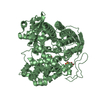
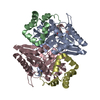
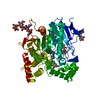
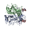
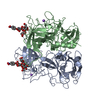
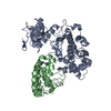
| Title | Structure of the C-terminal head domain of the fowl adenovirus type 4 fiber 1 | ||||||
 Components Components | Fiber-1 | ||||||
 Keywords Keywords | VIRAL PROTEIN / fiber / receptor binding | ||||||
| Function / homology | Fiber protein 1, C-terminal / Fiber protein, C-terminal domain superfamily / C-terminal head domain of the fowl adenovirus type 1 long fibre / Avian adenovirus fibre, N-terminal / Avian adenovirus fibre, N-terminal / virion attachment to host cell / Fiber-1 Function and homology information Function and homology information | ||||||
| Biological species |  Fowl adenovirus Fowl adenovirus | ||||||
| Method |  X-RAY DIFFRACTION / SYNCHROTRON / MOLECULAR REPLACEMENT / Resolution: 1.65 Å X-RAY DIFFRACTION / SYNCHROTRON / MOLECULAR REPLACEMENT / Resolution: 1.65 Å | ||||||
 Authors Authors | Song, Y.P. / Wei, Q. | ||||||
| Funding support |  China, 1items China, 1items
| ||||||
 Citation Citation | Journal: To Be Published Title: Structure of the C-terminal head domain of the fowl adenovirus type 4 fiber 1 Authors: Song, Y.P. / Wei, Q. | ||||||
| History |
|
- Structure visualization
| Structure viewer | Molecule: MolmilJmol/JSmol |
|---|
- Downloads & links
-Download
| PDBx/mmCIF format | 7x5t.cif.gz | 109.9 KB | Display | PDBx/mmCIF format |
|---|---|---|---|---|
| PDB format | pdb7x5t.ent.gz | Display | PDB format | |
| PDBx/mmJSON format | 7x5t.json.gz | Tree view | PDBx/mmJSON format | |
| Others |  Other downloads Other downloads |
-Validation report
| Arichive directory | https://data.pdbj.org/pub/pdb/validation_reports/x5/7x5tftp://data.pdbj.org/pub/pdb/validation_reports/x5/7x5t | HTTPS FTP |
|---|
-Related structure data
| Related structure data |  2iunS S: Starting model for refinement |
|---|---|
| Similar structure data |


-Links
 PDBj
PDBj- Assembly
| Deposited unit | 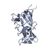
| ||||||||||||||||||
|---|---|---|---|---|---|---|---|---|---|---|---|---|---|---|---|---|---|---|---|
| 1 | 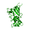
| ||||||||||||||||||
| Unit cell |
| ||||||||||||||||||
| Components on special symmetry positions |
|
-Components
| #1: Protein | Mass: 27264.137 Da / Num. of mol.: 1 Source method: isolated from a genetically manipulated source Source: (gene. exp.) Fowl adenovirus / Production host:  |
|---|---|
| #2: Water | ChemComp-HOH /  Mass: 18.015 Da / Num. of mol.: 215 / Source method: isolated from a natural source / Formula: H2O Mass: 18.015 Da / Num. of mol.: 215 / Source method: isolated from a natural source / Formula: H2O |
-Experimental details
-Experiment
| Experiment | Method: X-RAY DIFFRACTION / Number of used crystals: 1 |
|---|
- Sample preparation
| Crystal | Density Matthews: 2.42 Å3/Da / Density % sol: 49.23 % |
|---|---|
| Crystal grow | Temperature: 293 K / Method: evaporation Details: 0.1M Na citrate tribasic dihydrate pH5.0, 30% PEG monomethyl ether 550 |
-Data collection
| Diffraction | Mean temperature: 273 K / Serial crystal experiment: N |
|---|---|
| Diffraction source | Source: SYNCHROTRON / Site: ESRF  / Beamline: BM02 / Wavelength: 0.9793 Å / Beamline: BM02 / Wavelength: 0.9793 Å |
| Detector | Type: MARMOSAIC 225 mm CCD / Detector: CCD / Date: Dec 13, 2021 |
| Radiation | Protocol: SINGLE WAVELENGTH / Monochromatic (M) / Laue (L): M / Scattering type: x-ray |
| Radiation wavelength | Wavelength: 0.9793 Å / Relative weight: 1 |
| Reflection | Resolution: 1.65→47.65 Å / Num. obs: 33187 / % possible obs: 100 % / Redundancy: 75.7 % / Rmerge(I) obs: 0.128 / Net I/σ(I): 27.1 |
| Reflection shell | Resolution: 1.65→1.68 Å / Rmerge(I) obs: 3.295 / Num. unique obs: 1575 |
- Processing
| Software |
| |||||||||||||||||||||||||||||||||||||||||||||||||||||||||||||||||
|---|---|---|---|---|---|---|---|---|---|---|---|---|---|---|---|---|---|---|---|---|---|---|---|---|---|---|---|---|---|---|---|---|---|---|---|---|---|---|---|---|---|---|---|---|---|---|---|---|---|---|---|---|---|---|---|---|---|---|---|---|---|---|---|---|---|---|
| Refinement | Method to determine structure: MOLECULAR REPLACEMENT Starting model: 2IUN Resolution: 1.65→47.65 Å / Cor.coef. Fo:Fc: 0.982 / Cor.coef. Fo:Fc free: 0.961 / SU B: 3.672 / SU ML: 0.054 / Cross valid method: THROUGHOUT / σ(F): 0 / ESU R: 0.083 / ESU R Free: 0.078 / Stereochemistry target values: MAXIMUM LIKELIHOOD Details: HYDROGENS HAVE BEEN ADDED IN THE RIDING POSITIONS U VALUES : REFINED INDIVIDUALLY
| |||||||||||||||||||||||||||||||||||||||||||||||||||||||||||||||||
| Solvent computation | Ion probe radii: 0.8 Å / Shrinkage radii: 0.8 Å / VDW probe radii: 1.2 Å / Solvent model: MASK | |||||||||||||||||||||||||||||||||||||||||||||||||||||||||||||||||
| Displacement parameters | Biso max: 92.1 Å2 / Biso mean: 30.582 Å2 / Biso min: 18.72 Å2
| |||||||||||||||||||||||||||||||||||||||||||||||||||||||||||||||||
| Refinement step | Cycle: final / Resolution: 1.65→47.65 Å
| |||||||||||||||||||||||||||||||||||||||||||||||||||||||||||||||||
| Refine LS restraints |
| |||||||||||||||||||||||||||||||||||||||||||||||||||||||||||||||||
| LS refinement shell | Resolution: 1.65→1.693 Å / Rfactor Rfree error: 0 / Total num. of bins used: 20
|
