[English] 日本語
Yorodumi
- PDB-7x5b: Crystal structure of RuvB -

+
Open data


ID or keywords:

Loading...

-
Basic information

Entry
Database: PDB / ID: 7x5b
TitleCrystal structure of RuvB
ComponentsHolliday junction ATP-dependent DNA helicase RuvB
KeywordsMOTOR PROTEIN / RuvB / Holliday juncition / Homologous recombination / DNA damage repair / ATP hydrolysis
Function / homology
Function and homology information


cellular response to chromate / cellular response to selenite ion / Holliday junction resolvase complex / four-way junction helicase activity / four-way junction DNA binding / DNA recombination / DNA helicase / DNA repair / ATP hydrolysis activity / ATP binding / cytoplasm
Similarity search - Function
DNA helicase, Holliday junction RuvB-type / RuvB C-terminal winged helix domain / RuvB, AAA lid domain / RuvB C-terminal winged helix domain / RuvB AAA lid domain / RuvB-like P-loop domain / Holliday junction DNA helicase RuvB P-loop domain / Winged helix DNA-binding domain superfamily / Winged helix-like DNA-binding domain superfamily / ATPases associated with a variety of cellular activities ...DNA helicase, Holliday junction RuvB-type / RuvB C-terminal winged helix domain / RuvB, AAA lid domain / RuvB C-terminal winged helix domain / RuvB AAA lid domain / RuvB-like P-loop domain / Holliday junction DNA helicase RuvB P-loop domain / Winged helix DNA-binding domain superfamily / Winged helix-like DNA-binding domain superfamily / ATPases associated with a variety of cellular activities / AAA+ ATPase domain / P-loop containing nucleoside triphosphate hydrolase
Similarity search - Domain/homology
ADENOSINE-5'-DIPHOSPHATE / Holliday junction branch migration complex subunit RuvB
Similarity search - Component
Biological speciesPseudomonas aeruginosa PAO1 (bacteria)
MethodX-RAY DIFFRACTION / SYNCHROTRON / MOLECULAR REPLACEMENT / Resolution: 2.16 Å
AuthorsLin, Z. / Qu, Q. / Zhang, X. / Zhou, Z. / Dai, L.
Funding support China, 2items
OrganizationGrant numberCountry
National Natural Science Foundation of China (NSFC)31971222 China
National Natural Science Foundation of China (NSFC)32171194 China
CitationJournal: Front Plant Sci / Year: 2023
Title: Cryo-EM structure of the RuvAB-Holliday junction intermediate complex from .
Authors: Xu Zhang / Zixuan Zhou / Lin Dai / Yulin Chao / Zheng Liu / Mingdong Huang / Qianhui Qu / Zhonghui Lin /
Abstract: Holliday junction (HJ) is a four-way structured DNA intermediate in homologous recombination. In bacteria, the HJ-specific binding protein RuvA and the motor protein RuvB together form the RuvAB ...Holliday junction (HJ) is a four-way structured DNA intermediate in homologous recombination. In bacteria, the HJ-specific binding protein RuvA and the motor protein RuvB together form the RuvAB complex to catalyze HJ branch migration. (, Pa) is a ubiquitous opportunistic bacterial pathogen that can cause serious infection in a variety of host species, including vertebrate animals, insects and plants. Here, we describe the cryo-Electron Microscopy (cryo-EM) structure of the RuvAB-HJ intermediate complex from . The structure shows that two RuvA tetramers sandwich HJ at the junction center and disrupt base pairs at the branch points of RuvB-free HJ arms. Eight RuvB subunits are recruited by the RuvA octameric core and form two open-rings to encircle two opposite HJ arms. Each RuvB subunit individually binds a RuvA domain III. The four RuvB subunits within the ring display distinct subdomain conformations, and two of them engage the central DNA duplex at both strands with their C-terminal β-hairpins. Together with the biochemical analyses, our structure implicates a potential mechanism of RuvB motor assembly onto HJ DNA.
History
DepositionMar 4, 2022Deposition site: PDBJ / Processing site: PDBJ
Revision 1.0Mar 8, 2023Provider: repository / Type: Initial release
Revision 1.1Sep 20, 2023Group: Data collection / Database references
Category: chem_comp_atom / chem_comp_bond ...chem_comp_atom / chem_comp_bond / citation / citation_author
Item: _citation.country / _citation.journal_abbrev ..._citation.country / _citation.journal_abbrev / _citation.journal_id_CSD / _citation.journal_id_ISSN / _citation.journal_volume / _citation.page_first / _citation.page_last / _citation.pdbx_database_id_DOI / _citation.pdbx_database_id_PubMed / _citation.title / _citation.year
Revision 1.2Nov 29, 2023Group: Refinement description / Category: pdbx_initial_refinement_model

-
Structure visualization

Structure viewerMolecule:
MolmilJmol/JSmol

Downloads & links

-
Assembly

Deposited unit
A: Holliday junction ATP-dependent DNA helicase RuvB
hetero molecules


Theoretical massNumber of molelcules
Total (without water)39,3952
Polymers38,9681
Non-polymers4271
Water3,531196
1


  • Idetical with deposited unit
  • defined by author&software
TypeNameSymmetry operationNumber
identity operation1_555x,y,z1
Buried area800 Å2
ΔGint-5 kcal/mol
Surface area15570 Å2
MethodPISA
Unit cell
Length a, b, c (Å)85.677, 85.677, 76.973
Angle α, β, γ (deg.)90.000, 90.000, 120.000
Int Tables number170
Space group name H-MP65
Space group name HallP65
Symmetry operation#1: x,y,z
#2: x-y,x,z+5/6
#3: y,-x+y,z+1/6
#4: -y,x-y,z+2/3
#5: -x+y,-x,z+1/3
#6: -x,-y,z+1/2

-
Components

#1: Protein Holliday junction ATP-dependent DNA helicase RuvB


Mass: 38967.551 Da / Num. of mol.: 1 / Mutation: A146G
Source method: isolated from a genetically manipulated source
Source: (gene. exp.) Pseudomonas aeruginosa PAO1 (bacteria) / Strain: PAO1 / Gene: ruvB / Production host: Escherichia coli BL21(DE3) (bacteria) / Strain (production host): BL21(DE3) / References: UniProt: Q51426, DNA helicase
#2: Chemical ChemComp-ADP / ADENOSINE-5'-DIPHOSPHATE


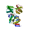
Mass: 427.201 Da / Num. of mol.: 1 / Source method: obtained synthetically / Formula: C10H15N5O10P2 / Feature type: SUBJECT OF INVESTIGATION / Comment: ADP, energy-carrying molecule*YM
#3: Water ChemComp-HOH / water


Mass: 18.015 Da / Num. of mol.: 196 / Source method: isolated from a natural source / Formula: H2O
Has ligand of interestY

-
Experimental details

-
Experiment

ExperimentMethod: X-RAY DIFFRACTION / Number of used crystals: 1

-
Sample preparation

CrystalDensity Matthews: 2.09 Å3/Da / Density % sol: 41.23 %
Crystal growTemperature: 291 K / Method: vapor diffusion, sitting drop
Details: 0.2 M LProline, 0.1 M HEPES pH 7.5, 10% w/v polyethylene glycol 3350, 10 mM Trimethylamine hydrochloride

-
Data collection

DiffractionMean temperature: 80 K / Serial crystal experiment: N
Diffraction sourceSource: SYNCHROTRON / Site: SSRF / Beamline: BL19U1 / Wavelength: 0.978 Å
DetectorType: DECTRIS PILATUS3 6M / Detector: PIXEL / Date: Jul 13, 2020
RadiationProtocol: SINGLE WAVELENGTH / Monochromatic (M) / Laue (L): M / Scattering type: x-ray
Radiation wavelengthWavelength: 0.978 Å / Relative weight: 1
ReflectionResolution: 2.1→50 Å / Num. obs: 17198 / % possible obs: 99.6 % / Redundancy: 20 % / Biso Wilson estimate: 22.37 Å2 / Rmerge(I) obs: 0.14 / Net I/σ(I): 23.9
Reflection shellResolution: 2.1→2.24 Å / CC1/2: 0.774

-
Processing

Software
NameVersionClassification
HKL-30001.13_2998data reduction
HKL-30001.13_2998data scaling
PHENIX1.19_4092refinement
MOLREPphasing
RefinementMethod to determine structure: MOLECULAR REPLACEMENT
Starting model: 6BLB
Resolution: 2.16→28.04 Å / SU ML: 0.1876 / Cross valid method: FREE R-VALUE / σ(F): 1.38 / Phase error: 20.9614 / Stereochemistry target values: GeoStd + Monomer Library
RfactorNum. reflection% reflection
Rfree0.2173 1717 9.98 %
Rwork0.1731 15481 -
obs0.1775 17198 99.65 %
Solvent computationShrinkage radii: 0.9 Å / VDW probe radii: 1.11 Å / Solvent model: FLAT BULK SOLVENT MODEL
Displacement parametersBiso mean: 27.14 Å2
Refinement stepCycle: LAST / Resolution: 2.16→28.04 Å
ProteinNucleic acidLigandSolventTotal
Num. atoms2440 0 27 196 2663
Refine LS restraints
Refine-IDTypeDev idealNumber
X-RAY DIFFRACTIONf_bond_d0.00692508
X-RAY DIFFRACTIONf_angle_d0.90823398
X-RAY DIFFRACTIONf_chiral_restr0.0544391
X-RAY DIFFRACTIONf_plane_restr0.0057443
X-RAY DIFFRACTIONf_dihedral_angle_d18.73921533
LS refinement shell
Resolution (Å)Rfactor RfreeNum. reflection RfreeRfactor RworkNum. reflection RworkRefine-ID% reflection obs (%)
2.16-2.230.25651400.19731242X-RAY DIFFRACTION96.58
2.23-2.30.25841420.19261280X-RAY DIFFRACTION99.86
2.3-2.380.23491430.18411303X-RAY DIFFRACTION100
2.38-2.470.24371420.18951273X-RAY DIFFRACTION100
2.47-2.590.23221460.18821291X-RAY DIFFRACTION100
2.59-2.720.27951430.19271278X-RAY DIFFRACTION100
2.72-2.890.19721440.18061296X-RAY DIFFRACTION100
2.89-3.120.22911380.17261302X-RAY DIFFRACTION100
3.12-3.430.18891430.17411301X-RAY DIFFRACTION100
3.43-3.930.21281500.16071287X-RAY DIFFRACTION100
3.93-4.940.17031430.14741297X-RAY DIFFRACTION99.93
4.94-28.040.22441430.17191331X-RAY DIFFRACTION99.59

+
About Yorodumi

-
News

-
Feb 9, 2022. New format data for meta-information of EMDB entries

New format data for meta-information of EMDB entries

  • Version 3 of the EMDB header file is now the official format.
  • The previous official version 1.9 will be removed from the archive.

Related info.:EMDB header

External links:wwPDB to switch to version 3 of the EMDB data model

-
Aug 12, 2020. Covid-19 info

Covid-19 info

URL: https://pdbjlvh1.pdbj.org/emnavi/covid19.php

New page: Covid-19 featured information page in EM Navigator.

Related info.:Covid-19 info / Mar 5, 2020. Novel coronavirus structure data

+
Mar 5, 2020. Novel coronavirus structure data

Novel coronavirus structure data

Related info.:Yorodumi Speices / Aug 12, 2020. Covid-19 info

External links:COVID-19 featured content - PDBj / Molecule of the Month (242):Coronavirus Proteases

+
Jan 31, 2019. EMDB accession codes are about to change! (news from PDBe EMDB page)

EMDB accession codes are about to change! (news from PDBe EMDB page)

  • The allocation of 4 digits for EMDB accession codes will soon come to an end. Whilst these codes will remain in use, new EMDB accession codes will include an additional digit and will expand incrementally as the available range of codes is exhausted. The current 4-digit format prefixed with “EMD-” (i.e. EMD-XXXX) will advance to a 5-digit format (i.e. EMD-XXXXX), and so on. It is currently estimated that the 4-digit codes will be depleted around Spring 2019, at which point the 5-digit format will come into force.
  • The EM Navigator/Yorodumi systems omit the EMD- prefix.

Related info.:Q: What is EMD? / ID/Accession-code notation in Yorodumi/EM Navigator

External links:EMDB Accession Codes are Changing Soon! / Contact to PDBj

+
Jul 12, 2017. Major update of PDB

Major update of PDB

  • wwPDB released updated PDB data conforming to the new PDBx/mmCIF dictionary.
  • This is a major update changing the version number from 4 to 5, and with Remediation, in which all the entries are updated.
  • In this update, many items about electron microscopy experimental information are reorganized (e.g. em_software).
  • Now, EM Navigator and Yorodumi are based on the updated data.

External links:wwPDB Remediation / Enriched Model Files Conforming to OneDep Data Standards Now Available in the PDB FTP Archive

-
Yorodumi

Thousand views of thousand structures

  • Yorodumi is a browser for structure data from EMDB, PDB, SASBDB, etc.
  • This page is also the successor to EM Navigator detail page, and also detail information page/front-end page for Omokage search.
  • The word "yorodu" (or yorozu) is an old Japanese word meaning "ten thousand". "mi" (miru) is to see.

Related info.:EMDB / PDB / SASBDB / Comparison of 3 databanks / Yorodumi Search / Aug 31, 2016. New EM Navigator & Yorodumi / Yorodumi Papers / Jmol/JSmol / Function and homology information / Changes in new EM Navigator and Yorodumi

Read more