 Movie
Movie Controller

[English] 日本語
 Yorodumi
- PDB-7x3z: Crystal structure of human 17beta-hydroxysteroid dehydrogenase ty... -
+ Open data

- Basic information
| Entry | Database: PDB / ID: 7x3z | ||||||
|---|---|---|---|---|---|---|---|
| Title | Crystal structure of human 17beta-hydroxysteroid dehydrogenase type 1 complexed with estrone and NAD | ||||||
 Components Components | 17-beta-hydroxysteroid dehydrogenase type 1 | ||||||
 Keywords Keywords | OXIDOREDUCTASE / 17BETA-HSD1 / ESTRONE / FUNCTIONAL ANALYSES / STEROID / NAD/NADH | ||||||
| Function / homology |  Function and homology information Function and homology information17-beta-hydroxysteroid dehydrogenase (NADP+) activity / cellular response to metal ion / estrogen biosynthetic process / 3(or 17)beta-hydroxysteroid dehydrogenase / estradiol binding / Estrogen biosynthesis / testosterone dehydrogenase (NADP+) activity / testosterone biosynthetic process / testosterone dehydrogenase (NAD+) activity / 17beta-estradiol 17-dehydrogenase ...17-beta-hydroxysteroid dehydrogenase (NADP+) activity / cellular response to metal ion / estrogen biosynthetic process / 3(or 17)beta-hydroxysteroid dehydrogenase / estradiol binding / Estrogen biosynthesis / testosterone dehydrogenase (NADP+) activity / testosterone biosynthetic process / testosterone dehydrogenase (NAD+) activity / 17beta-estradiol 17-dehydrogenase / estradiol 17-beta-dehydrogenase [NAD(P)+] activity / NADP+ binding / lysosome organization / The canonical retinoid cycle in rods (twilight vision) / small molecule binding / adipose tissue development / skeletal muscle tissue development / steroid binding / bone development / NADP binding / gene expression / protein homodimerization activity / cytosol Similarity search - Function | ||||||
| Biological species |  Homo sapiens (human) Homo sapiens (human) | ||||||
| Method |  X-RAY DIFFRACTION / SYNCHROTRON / MOLECULAR REPLACEMENT / molecular replacement / Resolution: 2.25 Å X-RAY DIFFRACTION / SYNCHROTRON / MOLECULAR REPLACEMENT / molecular replacement / Resolution: 2.25 Å | ||||||
| Model details | 17beta-HSD1-E1 complex | ||||||
 Authors Authors | Li, T. / Lin, S.X. / Yin, H. | ||||||
| Funding support | 1items
| ||||||
 Citation Citation | Journal: J.Steroid Biochem.Mol.Biol. / Year: 2023 Title: New insights into the substrate inhibition of human 17 beta-hydroxysteroid dehydrogenase type 1. Authors: Li, T. / Song, X. / Stephen, P. / Yin, H. / Lin, S.X. | ||||||
| History |
|
- Structure visualization
| Structure viewer | Molecule: MolmilJmol/JSmol |
|---|
- Downloads & links
-Download
| PDBx/mmCIF format | 7x3z.cif.gz | 123.7 KB | Display | PDBx/mmCIF format |
|---|---|---|---|---|
| PDB format | pdb7x3z.ent.gz | 93.5 KB | Display | PDB format |
| PDBx/mmJSON format | 7x3z.json.gz | Tree view | PDBx/mmJSON format | |
| Others |  Other downloads Other downloads |
-Validation report
| Arichive directory | https://data.pdbj.org/pub/pdb/validation_reports/x3/7x3zftp://data.pdbj.org/pub/pdb/validation_reports/x3/7x3z | HTTPS FTP |
|---|
-Related structure data
| Related structure data | 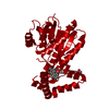 1jtvS S: Starting model for refinement |
|---|---|
| Similar structure data |
-Links
 PDBj
PDBj


- Assembly
| Deposited unit | 
| ||||||||
|---|---|---|---|---|---|---|---|---|---|
| 1 |
| ||||||||
| Unit cell |
|
-Components
| #1: Protein | Mass: 34989.000 Da / Num. of mol.: 2 Source method: isolated from a genetically manipulated source Source: (gene. exp.) Homo sapiens (human) / Gene: HSD17B1, E17KSR, EDH17B1, EDH17B2, EDHB17, SDR28C1 / Cell line (production host): Sf9 / Production host:   Spodoptera frugiperda (fall armyworm) Spodoptera frugiperda (fall armyworm)References: UniProt: P14061, 3(or 17)beta-hydroxysteroid dehydrogenase, 17beta-estradiol 17-dehydrogenase #2: Chemical |   Mass: 663.425 Da / Num. of mol.: 2 / Source method: obtained synthetically / Formula: C21H27N7O14P2 / Feature type: SUBJECT OF INVESTIGATION / Comment: NAD*YM Mass: 663.425 Da / Num. of mol.: 2 / Source method: obtained synthetically / Formula: C21H27N7O14P2 / Feature type: SUBJECT OF INVESTIGATION / Comment: NAD*YM#3: Chemical | ChemComp-J3Z / ( |   Mass: 270.366 Da / Num. of mol.: 1 / Source method: obtained synthetically / Formula: C18H22O2 / Feature type: SUBJECT OF INVESTIGATION Mass: 270.366 Da / Num. of mol.: 1 / Source method: obtained synthetically / Formula: C18H22O2 / Feature type: SUBJECT OF INVESTIGATION#4: Water | ChemComp-HOH / |  Mass: 18.015 Da / Num. of mol.: 77 / Source method: isolated from a natural source / Formula: H2O Mass: 18.015 Da / Num. of mol.: 77 / Source method: isolated from a natural source / Formula: H2OHas ligand of interest | Y | |
|---|
-Experimental details
-Experiment
| Experiment | Method: X-RAY DIFFRACTION / Number of used crystals: 1 |
|---|
- Sample preparation
| Crystal | Density Matthews: 1.95 Å3/Da / Density % sol: 36.82 % |
|---|---|
| Crystal grow | Temperature: 300 K / Method: vapor diffusion, hanging drop / Details: KH2PO4, PEG 8000 / PH range: 7.5-7.8 |
-Data collection
| Diffraction | Mean temperature: 100 K / Serial crystal experiment: N |
|---|---|
| Diffraction source | Source: SYNCHROTRON / Site: APS  / Beamline: 31-ID / Wavelength: 0.97931 Å / Beamline: 31-ID / Wavelength: 0.97931 Å |
| Detector | Type: MAR CCD 165 mm / Detector: CCD / Date: Feb 8, 2018 |
| Radiation | Protocol: SINGLE WAVELENGTH / Monochromatic (M) / Laue (L): M / Scattering type: x-ray |
| Radiation wavelength | Wavelength: 0.97931 Å / Relative weight: 1 |
| Reflection | Resolution: 2.25→25 Å / Num. obs: 26759 / % possible obs: 99.9 % / Redundancy: 7.3 % / CC1/2: 0.983 / Rpim(I) all: 0.075 / Rrim(I) all: 0.202 / Net I/σ(I): 7.9 / Num. measured all: 196005 |
| Reflection shell | Resolution: 2.25→2.37 Å / % possible obs: 100 % / Redundancy: 7.4 % / Num. measured all: 28323 / Num. unique obs: 3824 / CC1/2: 0.932 / Rpim(I) all: 0.185 / Rrim(I) all: 0.509 / Net I/σ(I) obs: 3.4 |
-Phasing
| Phasing | Method: molecular replacement | ||||||
|---|---|---|---|---|---|---|---|
| Phasing MR | R rigid body: 0.562
|
- Processing
| Software |
| ||||||||||||||||||||||||||||||||||||||||||||||||||||||||||||||||||||||||||||||||||||||||||||||||||||||||||||||||||||||||||||||||||||||||||||||||||||||||||||||||||||||||||||||||||||||
|---|---|---|---|---|---|---|---|---|---|---|---|---|---|---|---|---|---|---|---|---|---|---|---|---|---|---|---|---|---|---|---|---|---|---|---|---|---|---|---|---|---|---|---|---|---|---|---|---|---|---|---|---|---|---|---|---|---|---|---|---|---|---|---|---|---|---|---|---|---|---|---|---|---|---|---|---|---|---|---|---|---|---|---|---|---|---|---|---|---|---|---|---|---|---|---|---|---|---|---|---|---|---|---|---|---|---|---|---|---|---|---|---|---|---|---|---|---|---|---|---|---|---|---|---|---|---|---|---|---|---|---|---|---|---|---|---|---|---|---|---|---|---|---|---|---|---|---|---|---|---|---|---|---|---|---|---|---|---|---|---|---|---|---|---|---|---|---|---|---|---|---|---|---|---|---|---|---|---|---|---|---|---|---|
| Refinement | Method to determine structure: MOLECULAR REPLACEMENT Starting model: 1JTV Resolution: 2.25→24.99 Å / Cor.coef. Fo:Fc: 0.954 / Cor.coef. Fo:Fc free: 0.92 / SU B: 8.21 / SU ML: 0.197 / Cross valid method: THROUGHOUT / ESU R: 0.328 / ESU R Free: 0.243 / Stereochemistry target values: MAXIMUM LIKELIHOOD / Details: HYDROGENS HAVE BEEN ADDED IN THE RIDING POSITIONS
| ||||||||||||||||||||||||||||||||||||||||||||||||||||||||||||||||||||||||||||||||||||||||||||||||||||||||||||||||||||||||||||||||||||||||||||||||||||||||||||||||||||||||||||||||||||||
| Solvent computation | Ion probe radii: 0.8 Å / Shrinkage radii: 0.8 Å / VDW probe radii: 1.2 Å / Solvent model: MASK | ||||||||||||||||||||||||||||||||||||||||||||||||||||||||||||||||||||||||||||||||||||||||||||||||||||||||||||||||||||||||||||||||||||||||||||||||||||||||||||||||||||||||||||||||||||||
| Displacement parameters | Biso mean: 39.441 Å2
| ||||||||||||||||||||||||||||||||||||||||||||||||||||||||||||||||||||||||||||||||||||||||||||||||||||||||||||||||||||||||||||||||||||||||||||||||||||||||||||||||||||||||||||||||||||||
| Refinement step | Cycle: 1 / Resolution: 2.25→24.99 Å
| ||||||||||||||||||||||||||||||||||||||||||||||||||||||||||||||||||||||||||||||||||||||||||||||||||||||||||||||||||||||||||||||||||||||||||||||||||||||||||||||||||||||||||||||||||||||
| Refine LS restraints |
|