+ データを開く
- 基本情報
基本情報
| 登録情報 | データベース: PDB / ID: 7wqt | ||||||
|---|---|---|---|---|---|---|---|
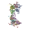
| タイトル | Cryo-EM structure of VWF D'D3 dimer complexed with D1D2 at 4.3 angstron resolution (VWF tube) | ||||||
 要素 要素 |
| ||||||
 キーワード キーワード | BLOOD CLOTTING / blood / VWF / von Willebrand factor / von Willebrand disease / blood coagulation / multimer assembly / VWF assembly / D'D3 domain / D1D2 domain / D'D3 dimer / D1D2 Dimer / VWF Tube / repeating unit | ||||||
| 機能・相同性 |  機能・相同性情報 機能・相同性情報Defective VWF binding to collagen type I / Enhanced cleavage of VWF variant by ADAMTS13 / Defective VWF cleavage by ADAMTS13 variant / Defective F8 binding to von Willebrand factor / Enhanced binding of GP1BA variant to VWF multimer:collagen / Defective binding of VWF variant to GPIb:IX:V / Weibel-Palade body / hemostasis / platelet alpha granule / Platelet Adhesion to exposed collagen ...Defective VWF binding to collagen type I / Enhanced cleavage of VWF variant by ADAMTS13 / Defective VWF cleavage by ADAMTS13 variant / Defective F8 binding to von Willebrand factor / Enhanced binding of GP1BA variant to VWF multimer:collagen / Defective binding of VWF variant to GPIb:IX:V / Weibel-Palade body / hemostasis / platelet alpha granule / Platelet Adhesion to exposed collagen / GP1b-IX-V activation signalling / p130Cas linkage to MAPK signaling for integrins / cell-substrate adhesion / Defective F8 cleavage by thrombin / Platelet Aggregation (Plug Formation) / GRB2:SOS provides linkage to MAPK signaling for Integrins / positive regulation of intracellular signal transduction / immunoglobulin binding / Integrin cell surface interactions / collagen binding / Intrinsic Pathway of Fibrin Clot Formation / Integrin signaling / extracellular matrix / platelet alpha granule lumen / Signaling by high-kinase activity BRAF mutants / MAP2K and MAPK activation / platelet activation / response to wounding / integrin binding / Signaling by RAF1 mutants / Signaling by moderate kinase activity BRAF mutants / Paradoxical activation of RAF signaling by kinase inactive BRAF / Signaling downstream of RAS mutants / blood coagulation / Signaling by BRAF and RAF1 fusions / Platelet degranulation / protein-folding chaperone binding / : / protease binding / cell adhesion / endoplasmic reticulum / extracellular space / extracellular exosome / extracellular region / identical protein binding 類似検索 - 分子機能 | ||||||
| 生物種 |  Homo sapiens (ヒト) Homo sapiens (ヒト) | ||||||
| 手法 | 電子顕微鏡法 / 単粒子再構成法 / クライオ電子顕微鏡法 / 解像度: 4.3 Å | ||||||
 データ登録者 データ登録者 | Zeng, J.W. / Shu, Z.M. / Zhou, A.W. | ||||||
| 資金援助 |  中国, 1件 中国, 1件
| ||||||
 引用 引用 |  ジャーナル: Blood / 年: 2022 ジャーナル: Blood / 年: 2022タイトル: Structural basis of von Willebrand factor multimerization and tubular storage. 著者: Jianwei Zeng / Zimei Shu / Qian Liang / Jing Zhang / Wenman Wu / Xuefeng Wang / Aiwu Zhou /  要旨: The von Willebrand factor (VWF) propeptide (domains D1D2) is essential for the assembly of VWF multimers and its tubular storage in Weibel-Palade bodies. However, detailed molecular mechanism ...The von Willebrand factor (VWF) propeptide (domains D1D2) is essential for the assembly of VWF multimers and its tubular storage in Weibel-Palade bodies. However, detailed molecular mechanism underlying this propeptide dependence is unclear. Here, we prepared Weibel-Palade body-like tubules using the N-terminal fragment of VWF and solved the cryo-electron microscopy structures of the tubule at atomic resolution. Detailed structural and biochemical analysis indicate that the propeptide forms a homodimer at acidic pH through the D2:D2 binding interface and then recruits 2 D'D3 domains, forming an intertwined D1D2D'D3 homodimer in essence. Stacking of these homodimers by the intermolecular D1:D2 interfaces brings 2 D3 domains face-to-face and facilitates their disulfide linkages and multimerization of VWF. Sequential stacking of these homodimers leads to a right-hand helical tubule for VWF storage. The clinically identified VWF mutations in the propeptide disrupted different steps of the assembling process, leading to diminished VWF multimers in von Willebrand diseases (VWD). Overall, these results indicate that the propeptide serves as a pH-sensing template for VWF multimerization and tubular storage. This sheds light on delivering normal propeptide as a template to rectify the defects in multimerization of VWD mutants. | ||||||
| 履歴 |
|
- 構造の表示
構造の表示
| 構造ビューア | 分子:  Molmil Molmil Jmol/JSmol Jmol/JSmol |
|---|
- ダウンロードとリンク
- ダウンロード
| PDBx/mmCIF形式 |  7wqt.cif.gz 7wqt.cif.gz | 3.1 MB | 表示 |  PDBx/mmCIF形式 PDBx/mmCIF形式 |
|---|---|---|---|---|
| PDB形式 |  pdb7wqt.ent.gz pdb7wqt.ent.gz | 表示 |  PDB形式 PDB形式 | |
| PDBx/mmJSON形式 |  7wqt.json.gz 7wqt.json.gz | ツリー表示 |  PDBx/mmJSON形式 PDBx/mmJSON形式 | |
| その他 |  その他のダウンロード その他のダウンロード |
-検証レポート
| 文書・要旨 |  7wqt_validation.pdf.gz 7wqt_validation.pdf.gz | 1.7 MB | 表示 |  wwPDB検証レポート wwPDB検証レポート |
|---|---|---|---|---|
| 文書・詳細版 |  7wqt_full_validation.pdf.gz 7wqt_full_validation.pdf.gz | 1.7 MB | 表示 | |
| XML形式データ |  7wqt_validation.xml.gz 7wqt_validation.xml.gz | 393.6 KB | 表示 | |
| CIF形式データ |  7wqt_validation.cif.gz 7wqt_validation.cif.gz | 651.6 KB | 表示 | |
| アーカイブディレクトリ |  https://data.pdbj.org/pub/pdb/validation_reports/wq/7wqt https://data.pdbj.org/pub/pdb/validation_reports/wq/7wqt ftp://data.pdbj.org/pub/pdb/validation_reports/wq/7wqt ftp://data.pdbj.org/pub/pdb/validation_reports/wq/7wqt | HTTPS FTP |
-関連構造データ
| 関連構造データ |  32713MC  7wppC  7wpqC  7wprC  7wpsC M: このデータのモデリングに利用したマップデータ C: 同じ文献を引用 ( |
|---|---|
| 類似構造データ | 類似検索 - 機能・相同性  F&H 検索 F&H 検索 |
- リンク
リンク
- 集合体
集合体
| 登録構造単位 | 
|
|---|---|
| 1 |
|
- 要素
要素
| #1: タンパク質 | 分子量: 81427.703 Da / 分子数: 16 / 断片: D1D2 domain / 由来タイプ: 組換発現 / 由来: (組換発現)  Homo sapiens (ヒト) / 遺伝子: VWF, F8VWF / 発現宿主: Homo sapiens (ヒト) / 遺伝子: VWF, F8VWF / 発現宿主:  Homo sapiens (ヒト) / 参照: UniProt: P04275 Homo sapiens (ヒト) / 参照: UniProt: P04275#2: タンパク質 | 分子量: 54232.809 Da / 分子数: 16 / 断片: D'D3 domain / 由来タイプ: 組換発現 / 由来: (組換発現)  Homo sapiens (ヒト) / 遺伝子: VWF, F8VWF / 発現宿主: Homo sapiens (ヒト) / 遺伝子: VWF, F8VWF / 発現宿主:  Homo sapiens (ヒト) / 参照: UniProt: P04275 Homo sapiens (ヒト) / 参照: UniProt: P04275#3: 糖 | ChemComp-NAG / #4: 化合物 | ChemComp-CA / 研究の焦点であるリガンドがあるか | Y | Has protein modification | Y | |
|---|
-実験情報
-実験
| 実験 | 手法: 電子顕微鏡法 |
|---|---|
| EM実験 | 試料の集合状態: PARTICLE / 3次元再構成法: 単粒子再構成法 |
- 試料調製
試料調製
| 構成要素 | 名称: VWF D'D3-D1D2 complex (8-9 units) / タイプ: COMPLEX / Entity ID: #2 / 由来: RECOMBINANT |
|---|---|
| 由来(天然) | 生物種:  Homo sapiens (ヒト) Homo sapiens (ヒト) |
| 由来(組換発現) | 生物種:  Homo sapiens (ヒト) Homo sapiens (ヒト) |
| 緩衝液 | pH: 7.4 |
| 試料 | 包埋: NO / シャドウイング: NO / 染色: NO / 凍結: YES |
| 急速凍結 | 凍結剤: ETHANE |
- 電子顕微鏡撮影
| 実験機器 |  モデル: Titan Krios / 画像提供: FEI Company |
|---|---|
| 顕微鏡 | モデル: FEI TITAN KRIOS |
| 電子銃 | 電子線源:  FIELD EMISSION GUN / 加速電圧: 300 kV / 照射モード: OTHER FIELD EMISSION GUN / 加速電圧: 300 kV / 照射モード: OTHER |
| 電子レンズ | モード: OTHER / 最大 デフォーカス(公称値): 2000 nm / 最小 デフォーカス(公称値): 1000 nm |
| 撮影 | 電子線照射量: 50 e/Å2 フィルム・検出器のモデル: GATAN K3 BIOQUANTUM (6k x 4k) |
- 解析
解析
| CTF補正 | タイプ: PHASE FLIPPING AND AMPLITUDE CORRECTION |
|---|---|
| 3次元再構成 | 解像度: 4.3 Å / 解像度の算出法: FSC 0.143 CUT-OFF / 粒子像の数: 220564 / 対称性のタイプ: POINT |
 ムービー
ムービー コントローラー


 PDBj
PDBj


