- PDB-7wdt: 6-sulfo-beta-D-N-acetylglucosaminidase from Bifidobacterium bifid... -
+
Open data
ID or keywords:
Loading...
-
Basic information
Entry
Database: PDB / ID: 7wdt
Title
6-sulfo-beta-D-N-acetylglucosaminidase from Bifidobacterium bifidum in complex with GlcNAc-6S
Components
Beta-N-acetylhexosaminidase
Keywords
HYDROLASE / Glycoside Hydrolase family 20
Function / homology
Function and homology information
glycosaminoglycan metabolic process / beta-N-acetylhexosaminidase activity / beta-N-acetylhexosaminidase / carbohydrate metabolic process / membrane / metal ion binding Similarity search - Function
Beta-hexosaminidase / Glycoside hydrolase family 20, catalytic domain / Glycosyl hydrolase family 20, catalytic domain / Chitobiase/beta-hexosaminidase domain 2-like / Beta-hexosaminidase, bacterial type, N-terminal / Glycosyl hydrolase family 20, domain 2 / Chitobiase; domain 2 / Beta-hexosaminidase-like, domain 2 / Coagulation factors 5/8 type C domain (FA58C) profile. / F5/8 type C domain ...Beta-hexosaminidase / Glycoside hydrolase family 20, catalytic domain / Glycosyl hydrolase family 20, catalytic domain / Chitobiase/beta-hexosaminidase domain 2-like / Beta-hexosaminidase, bacterial type, N-terminal / Glycosyl hydrolase family 20, domain 2 / Chitobiase; domain 2 / Beta-hexosaminidase-like, domain 2 / Coagulation factors 5/8 type C domain (FA58C) profile. / F5/8 type C domain / Coagulation factor 5/8 C-terminal domain / Galactose-binding domain-like / Galactose-binding-like domain superfamily / Glycosidases / Glycoside hydrolase superfamily / Jelly Rolls / TIM Barrel / Alpha-Beta Barrel / Sandwich / 2-Layer Sandwich / Mainly Beta / Alpha Beta Similarity search - Domain/homology
Method to determine structure: SAD / Resolution: 1.65→48.76 Å / Cor.coef. Fo:Fc: 0.969 / Cor.coef. Fo:Fc free: 0.96 / SU B: 2.294 / SU ML: 0.073 / Cross valid method: THROUGHOUT / σ(F): 0 / ESU R: 0.089 / ESU R Free: 0.089 / Stereochemistry target values: MAXIMUM LIKELIHOOD Details: HYDROGENS HAVE BEEN ADDED IN THE RIDING POSITIONS U VALUES : REFINED INDIVIDUALLY
Rfactor
Num. reflection
% reflection
Selection details
Rfree
0.1998
5253
5 %
RANDOM
Rwork
0.1697
-
-
-
obs
0.1712
99053
99.45 %
-
Solvent computation
Ion probe radii: 0.8 Å / Shrinkage radii: 0.8 Å / VDW probe radii: 1.2 Å / Solvent model: MASK
In the structure databanks used in Yorodumi, some data are registered as the other names, "COVID-19 virus" and "2019-nCoV". Here are the details of the virus and the list of structure data.
Jan 31, 2019. EMDB accession codes are about to change! (news from PDBe EMDB page)
EMDB accession codes are about to change! (news from PDBe EMDB page)
The allocation of 4 digits for EMDB accession codes will soon come to an end. Whilst these codes will remain in use, new EMDB accession codes will include an additional digit and will expand incrementally as the available range of codes is exhausted. The current 4-digit format prefixed with “EMD-” (i.e. EMD-XXXX) will advance to a 5-digit format (i.e. EMD-XXXXX), and so on. It is currently estimated that the 4-digit codes will be depleted around Spring 2019, at which point the 5-digit format will come into force.
The EM Navigator/Yorodumi systems omit the EMD- prefix.
Related info.:Q: What is EMD? / ID/Accession-code notation in Yorodumi/EM Navigator
Yorodumi is a browser for structure data from EMDB, PDB, SASBDB, etc.
This page is also the successor to EM Navigator detail page, and also detail information page/front-end page for Omokage search.
The word "yorodu" (or yorozu) is an old Japanese word meaning "ten thousand". "mi" (miru) is to see.
Related info.:EMDB / PDB / SASBDB / Comparison of 3 databanks / Yorodumi Search / Aug 31, 2016. New EM Navigator & Yorodumi / Yorodumi Papers / Jmol/JSmol / Function and homology information / Changes in new EM Navigator and Yorodumi
 Movie
Movie Controller

 Yorodumi
 Open data
 Basic information
 Components
 Keywords
 Function and homology information
 Bifidobacterium bifidum JCM 1254 (bacteria)
 X-RAY DIFFRACTION /
  Authors
 Japan, 2items
  Citation
 Structure visualization
 Downloads & links
 Other downloads
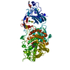
 PDBj
PDBj

 Assembly
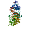
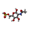
 Type: D-saccharide, beta linking / Mass: 301.271 Da / Num. of mol.: 1 / Source method: obtained synthetically / Formula: C8H15NO9S / Feature type: SUBJECT OF INVESTIGATION
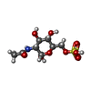
 Type: D-saccharide, alpha linking / Mass: 301.271 Da / Num. of mol.: 1 / Source method: obtained synthetically / Formula: C8H15NO9S / Feature type: SUBJECT OF INVESTIGATION
 Mass: 40.078 Da / Num. of mol.: 1 / Source method: obtained synthetically / Formula: Ca
 Mass: 18.015 Da / Num. of mol.: 619 / Source method: isolated from a natural source / Formula: H2O
 Sample preparation
 Processing