 Movie
Movie Controller


+ Open data

- Basic information
| Entry | Database: PDB / ID: 7wc2 | ||||||
|---|---|---|---|---|---|---|---|
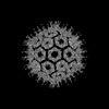
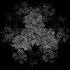
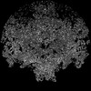
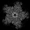
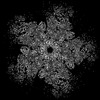
| Title | Cryo-EM structure of alphavirus, Getah virus | ||||||
 Components Components |
| ||||||
 Keywords Keywords | VIRUS / alphavirus / Getah virus / Togaviridae / structural genomics | ||||||
| Function / homology |  Function and homology information Function and homology informationtogavirin / T=4 icosahedral viral capsid / symbiont-mediated suppression of host toll-like receptor signaling pathway / host cell cytoplasm / serine-type endopeptidase activity / fusion of virus membrane with host endosome membrane / symbiont entry into host cell / virion attachment to host cell / host cell nucleus / host cell plasma membrane ...togavirin / T=4 icosahedral viral capsid / symbiont-mediated suppression of host toll-like receptor signaling pathway / host cell cytoplasm / serine-type endopeptidase activity / fusion of virus membrane with host endosome membrane / symbiont entry into host cell / virion attachment to host cell / host cell nucleus / host cell plasma membrane / virion membrane / structural molecule activity / proteolysis / RNA binding / membrane Similarity search - Function | ||||||
| Biological species |  Getah virus Getah virus | ||||||
| Method | ELECTRON MICROSCOPY / single particle reconstruction / cryo EM / Resolution: 3.5 Å | ||||||
 Authors Authors | Wang, M. / Sun, Z.Z. / Wang, J.F. | ||||||
| Funding support | 1items
| ||||||
 Citation Citation | Journal: To Be Published Title: Implications for the pathogenicity and antigenicity of alpha viruses revealed by a 3.5 angstrom Cryo-EM structure of Getah virus Authors: Wang, M. / Sun, Z.Z. / Wang, J.F. | ||||||
| History |
|
- Structure visualization
| Structure viewer | Molecule: MolmilJmol/JSmol |
|---|
- Downloads & links
-Download
| PDBx/mmCIF format | 7wc2.cif.gz | 600.2 KB | Display | PDBx/mmCIF format |
|---|---|---|---|---|
| PDB format | pdb7wc2.ent.gz | 497.7 KB | Display | PDB format |
| PDBx/mmJSON format | 7wc2.json.gz | Tree view | PDBx/mmJSON format | |
| Others |  Other downloads Other downloads |
-Validation report
| Summary document | 7wc2_validation.pdf.gz | 1.9 MB | Display | wwPDB validaton report |
|---|---|---|---|---|
| Full document | 7wc2_full_validation.pdf.gz | 2 MB | Display | |
| Data in XML | 7wc2_validation.xml.gz | 93.5 KB | Display | |
| Data in CIF | 7wc2_validation.cif.gz | 142.6 KB | Display | |
| Arichive directory | https://data.pdbj.org/pub/pdb/validation_reports/wc/7wc2ftp://data.pdbj.org/pub/pdb/validation_reports/wc/7wc2 | HTTPS FTP |
-Related structure data
| Related structure data |  32412MC 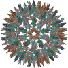 7vgaC 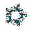 7wcoC M: map data used to model this data C: citing same article ( |
|---|---|
| Similar structure data |
-Links
 PDBj
PDBj


- Assembly
| Deposited unit | 
|
|---|---|
| 1 | x 60
|
| 2 |
|
| 3 | x 5
|
| 4 | x 6
|
| 5 | 
|
| Symmetry | Point symmetry: (Schoenflies symbol: I (icosahedral)) |
-Components
| #1: Protein | Mass: 47620.770 Da / Num. of mol.: 4 / Source method: isolated from a natural source / Source: (natural) Getah virus / References: UniProt: Q5Y388#2: Protein | Mass: 46416.793 Da / Num. of mol.: 4 / Source method: isolated from a natural source / Source: (natural) Getah virus / References: UniProt: Q5Y388#3: Polysaccharide | alpha-D-mannopyranose-(1-3)-[alpha-D-mannopyranose-(1-6)]beta-D-mannopyranose-(1-4)-2-acetamido-2- ...alpha-D-mannopyranose-(1-3)-[alpha-D-mannopyranose-(1-6)]beta-D-mannopyranose-(1-4)-2-acetamido-2-deoxy-beta-D-glucopyranose-(1-4)-2-acetamido-2-deoxy-beta-D-glucopyranose Source method: isolated from a genetically manipulated source Has ligand of interest | N | Has protein modification | Y | |
|---|
-Experimental details
-Experiment
| Experiment | Method: ELECTRON MICROSCOPY |
|---|---|
| EM experiment | Aggregation state: CELL / 3D reconstruction method: single particle reconstruction |
- Sample preparation
| Component | Name: Getah virus / Type: VIRUS / Entity ID: #1-#2 / Source: NATURAL |
|---|---|
| Source (natural) | Organism: Getah virus |
| Details of virus | Empty: NO / Enveloped: YES / Isolate: STRAIN / Type: VIRION |
| Natural host | Organism: Getah virus |
| Buffer solution | pH: 7.4 |
| Specimen | Embedding applied: NO / Shadowing applied: NO / Staining applied: NO / Vitrification applied: YES |
| Vitrification | Cryogen name: NITROGEN |
- Electron microscopy imaging
| Experimental equipment |  Model: Titan Krios / Image courtesy: FEI Company |
|---|---|
| Microscopy | Model: FEI TITAN KRIOS |
| Electron gun | Electron source:  FIELD EMISSION GUN / Accelerating voltage: 300 kV / Illumination mode: FLOOD BEAM FIELD EMISSION GUN / Accelerating voltage: 300 kV / Illumination mode: FLOOD BEAM |
| Electron lens | Mode: BRIGHT FIELD / Nominal defocus max: 2500 nm / Nominal defocus min: 1000 nm |
| Image recording | Electron dose: 35 e/Å2 / Film or detector model: GATAN K2 SUMMIT (4k x 4k) |
- Processing
| CTF correction | Type: PHASE FLIPPING ONLY |
|---|---|
| Particle selection | Num. of particles selected: 36066 / Details: region for particle picking |
| 3D reconstruction | Resolution: 3.5 Å / Resolution method: FSC 0.143 CUT-OFF / Num. of particles: 30996 / Symmetry type: POINT |
