 Movie
Movie Controller

[English] 日本語
 Yorodumi
- PDB-7w2w: Crystal structure of TxGH116 R786K mutant from Thermoanaerobacter... -
+ Open data

- Basic information
| Entry | Database: PDB / ID: 7w2w | ||||||||||||||||||
|---|---|---|---|---|---|---|---|---|---|---|---|---|---|---|---|---|---|---|---|
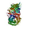
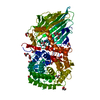
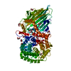
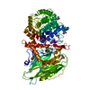
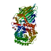
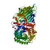
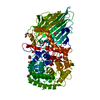
| Title | Crystal structure of TxGH116 R786K mutant from Thermoanaerobacterium xylanolyticum with glucose | ||||||||||||||||||
 Components Components | Glucosylceramidase | ||||||||||||||||||
 Keywords Keywords | HYDROLASE / TxGH116 / beta-glucosidase / mutant / Thermoanaerobacterium xylanolyticum / glucose | ||||||||||||||||||
| Function / homology |  Function and homology information Function and homology informationglucosylceramidase / glucosylceramide catabolic process / glucosylceramidase activity / beta-glucosidase activity / carbohydrate metabolic process / membrane / metal ion binding Similarity search - Function | ||||||||||||||||||
| Biological species |  Thermoanaerobacterium xylanolyticum (bacteria) Thermoanaerobacterium xylanolyticum (bacteria) | ||||||||||||||||||
| Method |  X-RAY DIFFRACTION / SYNCHROTRON / MOLECULAR REPLACEMENT / Resolution: 1.79 Å X-RAY DIFFRACTION / SYNCHROTRON / MOLECULAR REPLACEMENT / Resolution: 1.79 Å | ||||||||||||||||||
 Authors Authors | Huang, M. / Pengthaisong, S. / Charoenwattanasatien, R. / Jitonnom, J. / Ketudat Cairns, J.R. | ||||||||||||||||||
| Funding support |  Thailand, 5items Thailand, 5items
| ||||||||||||||||||
 Citation Citation | Journal: Catalysts / Year: 2022 Title: Systematic Functional and Computational Analysis of Glucose-Binding Residues in Glycoside Hydrolase Family GH116. Authors: Huang, M. / Pengthaisong, S. / Charoenwattanasatien, R. / Thinkumrob, N. / Jitonnom, J. / Ketudat Cairns, J.R. | ||||||||||||||||||
| History |
|
- Structure visualization
| Structure viewer | Molecule: MolmilJmol/JSmol |
|---|
- Downloads & links
-Download
| PDBx/mmCIF format | 7w2w.cif.gz | 186.4 KB | Display | PDBx/mmCIF format |
|---|---|---|---|---|
| PDB format | pdb7w2w.ent.gz | 142.3 KB | Display | PDB format |
| PDBx/mmJSON format | 7w2w.json.gz | Tree view | PDBx/mmJSON format | |
| Others |  Other downloads Other downloads |
-Validation report
| Arichive directory | https://data.pdbj.org/pub/pdb/validation_reports/w2/7w2wftp://data.pdbj.org/pub/pdb/validation_reports/w2/7w2w | HTTPS FTP |
|---|
-Related structure data
| Related structure data | 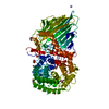 7w2sC 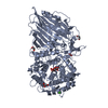 7w2tC 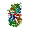 7w2vC 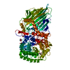 7w2xC 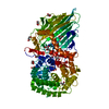 5bvuS S: Starting model for refinement C: citing same article ( |
|---|---|
| Similar structure data |


-Links
 PDBj
PDBj


- Assembly
| Deposited unit | 
| ||||||||
|---|---|---|---|---|---|---|---|---|---|
| 1 |
| ||||||||
| Unit cell |
|
-Components
-Protein , 1 types, 1 molecules A
| #1: Protein | Mass: 96564.422 Da / Num. of mol.: 1 / Mutation: R786K Source method: isolated from a genetically manipulated source Source: (gene. exp.) Thermoanaerobacterium xylanolyticum (strain ATCC 49914 / DSM 7097 / LX-11) (bacteria)Strain: ATCC 49914 / DSM 7097 / LX-11 / Gene: Thexy_2211 / Plasmid: pET30a / Production host: |
|---|
-Sugars , 2 types, 2 molecules 

| #2: Sugar | ChemComp-GLC /  Type: D-saccharide, alpha linking / Mass: 180.156 Da / Num. of mol.: 1 / Source method: obtained synthetically / Formula: C6H12O6 / Feature type: SUBJECT OF INVESTIGATION Type: D-saccharide, alpha linking / Mass: 180.156 Da / Num. of mol.: 1 / Source method: obtained synthetically / Formula: C6H12O6 / Feature type: SUBJECT OF INVESTIGATION |
|---|---|
| #3: Sugar | ChemComp-BGC /  Type: D-saccharide, beta linking / Mass: 180.156 Da / Num. of mol.: 1 / Source method: obtained synthetically / Formula: C6H12O6 / Feature type: SUBJECT OF INVESTIGATION Type: D-saccharide, beta linking / Mass: 180.156 Da / Num. of mol.: 1 / Source method: obtained synthetically / Formula: C6H12O6 / Feature type: SUBJECT OF INVESTIGATION |
-Non-polymers , 5 types, 478 molecules 


| #4: Chemical | ChemComp-GOL /  Mass: 92.094 Da / Num. of mol.: 12 / Source method: obtained synthetically / Formula: C3H8O3 / Feature type: SUBJECT OF INVESTIGATION Mass: 92.094 Da / Num. of mol.: 12 / Source method: obtained synthetically / Formula: C3H8O3 / Feature type: SUBJECT OF INVESTIGATION#5: Chemical | ChemComp-SO4 / |  Mass: 96.063 Da / Num. of mol.: 1 / Source method: obtained synthetically / Formula: SO4 Mass: 96.063 Da / Num. of mol.: 1 / Source method: obtained synthetically / Formula: SO4#6: Chemical | ChemComp-EDO / |  Mass: 62.068 Da / Num. of mol.: 1 / Source method: obtained synthetically / Formula: C2H6O2 Mass: 62.068 Da / Num. of mol.: 1 / Source method: obtained synthetically / Formula: C2H6O2#7: Chemical | ChemComp-CA / |  Mass: 40.078 Da / Num. of mol.: 1 / Source method: obtained synthetically / Formula: Ca Mass: 40.078 Da / Num. of mol.: 1 / Source method: obtained synthetically / Formula: Ca#8: Water | ChemComp-HOH / | Mass: 18.015 Da / Num. of mol.: 463 / Source method: isolated from a natural source / Formula: H2O |
|---|
-Details
| Has ligand of interest | Y |
|---|
-Experimental details
-Experiment
| Experiment | Method: X-RAY DIFFRACTION / Number of used crystals: 1 |
|---|
- Sample preparation
| Crystal | Density Matthews: 2.11 Å3/Da / Density % sol: 41.77 % |
|---|---|
| Crystal grow | Temperature: 288 K / Method: vapor diffusion, hanging drop / pH: 5.5 Details: 0.2 M AMMONIUM SULFATE, 22% PEG 3000, 0.1 M MES, PH 5.5 |
-Data collection
| Diffraction | Mean temperature: 100 K / Serial crystal experiment: N |
|---|---|
| Diffraction source | Source: SYNCHROTRON / Site: NSRRC  / Beamline: BL13B1 / Wavelength: 1 Å / Beamline: BL13B1 / Wavelength: 1 Å |
| Detector | Type: ADSC QUANTUM 315r / Detector: CCD / Date: Jul 10, 2020 |
| Radiation | Protocol: SINGLE WAVELENGTH / Monochromatic (M) / Laue (L): M / Scattering type: x-ray |
| Radiation wavelength | Wavelength: 1 Å / Relative weight: 1 |
| Reflection | Resolution: 1.79→50.01 Å / Num. obs: 75984 / % possible obs: 98.5 % / Redundancy: 7.2 % / Rmerge(I) obs: 0.121 / Net I/σ(I): 16.3 |
| Reflection shell | Resolution: 1.79→1.85 Å / Redundancy: 5.8 % / Rmerge(I) obs: 0.81 / Mean I/σ(I) obs: 1.9 / Num. unique obs: 6616 / CC1/2: 0.708 / % possible all: 86.7 |
- Processing
| Software |
| ||||||||||||||||||||||||||||||||||||||||||||||||||||||||||||
|---|---|---|---|---|---|---|---|---|---|---|---|---|---|---|---|---|---|---|---|---|---|---|---|---|---|---|---|---|---|---|---|---|---|---|---|---|---|---|---|---|---|---|---|---|---|---|---|---|---|---|---|---|---|---|---|---|---|---|---|---|---|
| Refinement | Method to determine structure: MOLECULAR REPLACEMENT Starting model: 5BVU Resolution: 1.79→50.01 Å / Cor.coef. Fo:Fc: 0.969 / Cor.coef. Fo:Fc free: 0.957 / SU B: 2.379 / SU ML: 0.073 / Cross valid method: THROUGHOUT / σ(F): 0 / ESU R: 0.111 / ESU R Free: 0.107 / Stereochemistry target values: MAXIMUM LIKELIHOOD Details: HYDROGENS HAVE BEEN ADDED IN THE RIDING POSITIONS U VALUES : REFINED INDIVIDUALLY
| ||||||||||||||||||||||||||||||||||||||||||||||||||||||||||||
| Solvent computation | Ion probe radii: 0.8 Å / Shrinkage radii: 0.8 Å / VDW probe radii: 1.2 Å / Solvent model: MASK | ||||||||||||||||||||||||||||||||||||||||||||||||||||||||||||
| Displacement parameters | Biso max: 74.19 Å2 / Biso mean: 22.193 Å2 / Biso min: 6.64 Å2
| ||||||||||||||||||||||||||||||||||||||||||||||||||||||||||||
| Refinement step | Cycle: final / Resolution: 1.79→50.01 Å
| ||||||||||||||||||||||||||||||||||||||||||||||||||||||||||||
| Refine LS restraints |
| ||||||||||||||||||||||||||||||||||||||||||||||||||||||||||||
| LS refinement shell | Resolution: 1.79→1.836 Å / Rfactor Rfree error: 0 / Total num. of bins used: 20
|
