 Movie
Movie Controller

[English] 日本語
 Yorodumi
- PDB-7vyw: Crystal structure of the chromodomain of Arabidopsis LHP1 in comp... -
+ Open data

- Basic information
| Entry | Database: PDB / ID: 7vyw | ||||||
|---|---|---|---|---|---|---|---|
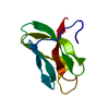
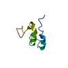
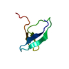
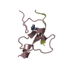
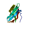
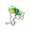
| Title | Crystal structure of the chromodomain of Arabidopsis LHP1 in complex with methylated histone H3K9 peptide | ||||||
 Components Components |
| ||||||
 Keywords Keywords | TRANSCRIPTION | ||||||
| Function / homology |  Function and homology information Function and homology informationvernalization response / shoot system morphogenesis / photoperiodism / GTP cyclohydrolase II activity / multidimensional cell growth / negative regulation of flower development / photoperiodism, flowering / flower development / negative regulation of gene expression, epigenetic / : ...vernalization response / shoot system morphogenesis / photoperiodism / GTP cyclohydrolase II activity / multidimensional cell growth / negative regulation of flower development / photoperiodism, flowering / flower development / negative regulation of gene expression, epigenetic / : / heterochromatin / chloroplast / euchromatin / heterochromatin formation / chromatin organization / sequence-specific DNA binding / cell differentiation / negative regulation of DNA-templated transcription / chromatin binding / DNA binding / identical protein binding / nucleus Similarity search - Function | ||||||
| Biological species |  | ||||||
| Method |  X-RAY DIFFRACTION / SYNCHROTRON / MOLECULAR REPLACEMENT / Resolution: 1.6 Å X-RAY DIFFRACTION / SYNCHROTRON / MOLECULAR REPLACEMENT / Resolution: 1.6 Å | ||||||
 Authors Authors | Liu, Y. / Zhang, M. / Min, J. | ||||||
| Funding support |  China, 1items China, 1items
| ||||||
 Citation Citation | Journal: J.Biol.Chem. / Year: 2022 Title: Structural basis for the recognition of methylated histone H3 by the Arabidopsis LHP1 chromodomain. Authors: Liu, Y. / Yang, X. / Zhou, M. / Yang, Y. / Li, F. / Yan, X. / Zhang, M. / Wei, Z. / Qin, S. / Min, J. | ||||||
| History |
|
- Structure visualization
| Structure viewer | Molecule: MolmilJmol/JSmol |
|---|
- Downloads & links
-Download
| PDBx/mmCIF format | 7vyw.cif.gz | 28.2 KB | Display | PDBx/mmCIF format |
|---|---|---|---|---|
| PDB format | pdb7vyw.ent.gz | 15.4 KB | Display | PDB format |
| PDBx/mmJSON format | 7vyw.json.gz | Tree view | PDBx/mmJSON format | |
| Others |  Other downloads Other downloads |
-Validation report
| Arichive directory | https://data.pdbj.org/pub/pdb/validation_reports/vy/7vywftp://data.pdbj.org/pub/pdb/validation_reports/vy/7vyw | HTTPS FTP |
|---|
-Related structure data
| Related structure data |  7vz2C  1q3lS S: Starting model for refinement C: citing same article ( |
|---|---|
| Similar structure data |


-Links
 PDBj
PDBj- Assembly
| Deposited unit | 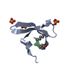
| ||||||||
|---|---|---|---|---|---|---|---|---|---|
| 1 |
| ||||||||
| Unit cell |
|
-Components
| #1: Protein | Mass: 6574.503 Da / Num. of mol.: 1 Source method: isolated from a genetically manipulated source Source: (gene. exp.)  | ||||
|---|---|---|---|---|---|
| #2: Protein/peptide | Mass: 716.827 Da / Num. of mol.: 1 / Source method: obtained synthetically / Source: (synth.) | ||||
| #3: Chemical |   Mass: 96.063 Da / Num. of mol.: 3 / Source method: isolated from a natural source / Formula: SO4 Mass: 96.063 Da / Num. of mol.: 3 / Source method: isolated from a natural source / Formula: SO4#4: Water | ChemComp-HOH / |  Mass: 18.015 Da / Num. of mol.: 59 / Source method: isolated from a natural source / Formula: H2O Mass: 18.015 Da / Num. of mol.: 59 / Source method: isolated from a natural source / Formula: H2OHas ligand of interest | Y | |
-Experimental details
-Experiment
| Experiment | Method: X-RAY DIFFRACTION / Number of used crystals: 1 |
|---|
- Sample preparation
| Crystal | Density Matthews: 1.75 Å3/Da / Density % sol: 29.66 % |
|---|---|
| Crystal grow | Temperature: 293 K / Method: vapor diffusion, sitting drop / Details: 0.1 M Hepes, pH 7.5, 2.0 M Ammonium sulfate |
-Data collection
| Diffraction | Mean temperature: 100 K / Serial crystal experiment: N |
|---|---|
| Diffraction source | Source: SYNCHROTRON / Site: SSRF / Beamline: BL17U1 / Wavelength: 1.5418 Å |
| Detector | Type: ADSC QUANTUM 315r / Detector: CCD / Date: Mar 3, 2019 |
| Radiation | Protocol: SINGLE WAVELENGTH / Monochromatic (M) / Laue (L): M / Scattering type: x-ray |
| Radiation wavelength | Wavelength: 1.5418 Å / Relative weight: 1 |
| Reflection | Resolution: 1.6→35.26 Å / Num. obs: 6673 / % possible obs: 90.29 % / Redundancy: 4.6 % / Biso Wilson estimate: 10.57 Å2 / CC1/2: 0.982 / Net I/σ(I): 23.2 |
| Reflection shell | Resolution: 1.6→1.66 Å / Num. unique obs: 294 / CC1/2: 0.968 |
- Processing
| Software |
| ||||||||||||||||||||||||
|---|---|---|---|---|---|---|---|---|---|---|---|---|---|---|---|---|---|---|---|---|---|---|---|---|---|
| Refinement | Method to determine structure: MOLECULAR REPLACEMENT Starting model: 1Q3L Resolution: 1.6→35.26 Å / SU ML: 0.15 / Cross valid method: THROUGHOUT / σ(F): 1.52 / Phase error: 21.17 / Stereochemistry target values: ML
| ||||||||||||||||||||||||
| Solvent computation | Shrinkage radii: 0.9 Å / VDW probe radii: 1.11 Å / Solvent model: FLAT BULK SOLVENT MODEL | ||||||||||||||||||||||||
| Displacement parameters | Biso max: 48.2 Å2 / Biso mean: 9.8733 Å2 / Biso min: 1.8 Å2 | ||||||||||||||||||||||||
| Refinement step | Cycle: final / Resolution: 1.6→35.26 Å
| ||||||||||||||||||||||||
| LS refinement shell | Refine-ID: X-RAY DIFFRACTION / Rfactor Rfree error: 0 / Total num. of bins used: 2
|
