 Movie
Movie Controller


+ Open data

- Basic information
| Entry | Database: PDB / ID: 7vrm | ||||||
|---|---|---|---|---|---|---|---|
| Title | crystal structure of BRD2-BD2 in complex with purine derivative | ||||||
 Components Components | Bromodomain-containing protein 2 | ||||||
 Keywords Keywords | TRANSCRIPTION/INHIBITOR / BET family / BET inhibitor / Bromodomain Inhibitor / BRD2-BD1 inhibitor / TRANSCRIPTION-INHIBITOR COMPLEX | ||||||
| Function / homology |  Function and homology information Function and homology informationacetylation-dependent protein binding / histone H3K14ac reader activity / chromatin looping / histone H4K5ac reader activity / histone H4K12ac reader activity / RUNX3 regulates p14-ARF / positive regulation of T-helper 17 cell lineage commitment / protein localization to chromatin / neural tube closure / nucleosome assembly ...acetylation-dependent protein binding / histone H3K14ac reader activity / chromatin looping / histone H4K5ac reader activity / histone H4K12ac reader activity / RUNX3 regulates p14-ARF / positive regulation of T-helper 17 cell lineage commitment / protein localization to chromatin / neural tube closure / nucleosome assembly / spermatogenesis / histone binding / nuclear speck / chromatin remodeling / protein serine/threonine kinase activity / chromatin binding / regulation of transcription by RNA polymerase II / chromatin / nucleoplasm / nucleus / cytoplasm Similarity search - Function | ||||||
| Biological species |  Homo sapiens (human) Homo sapiens (human) | ||||||
| Method |  X-RAY DIFFRACTION / MOLECULAR REPLACEMENT / Resolution: 1.1 Å X-RAY DIFFRACTION / MOLECULAR REPLACEMENT / Resolution: 1.1 Å | ||||||
 Authors Authors | Padmanabhan, B. / Arole, A. / Deshmukh, P. / Ashok, S. / Mathur, S. | ||||||
| Funding support | 1items
| ||||||
 Citation Citation | Journal: Acta Crystallogr D Struct Biol / Year: 2023 Title: Structural and biochemical insights into purine-based drug molecules in hBRD2 delineate a unique binding mode opening new vistas in the design of inhibitors of the BET family. Authors: Arole, A.H. / Deshmukh, P. / Sridhar, A. / Mathur, S. / Mahalingaswamy, M. / Subramanya, H. / Dalavaikodihalli Nanjaiah, N. / Padmanabhan, B. | ||||||
| History |
|
- Structure visualization
| Structure viewer | Molecule: MolmilJmol/JSmol |
|---|
- Downloads & links
-Download
| PDBx/mmCIF format | 7vrm.cif.gz | 69.6 KB | Display | PDBx/mmCIF format |
|---|---|---|---|---|
| PDB format | pdb7vrm.ent.gz | 48.9 KB | Display | PDB format |
| PDBx/mmJSON format | 7vrm.json.gz | Tree view | PDBx/mmJSON format | |
| Others |  Other downloads Other downloads |
-Validation report
| Arichive directory | https://data.pdbj.org/pub/pdb/validation_reports/vr/7vrmftp://data.pdbj.org/pub/pdb/validation_reports/vr/7vrm | HTTPS FTP |
|---|
-Related structure data
| Related structure data |  7vrhC 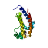 7vriC 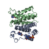 7vrkC  7vroC 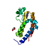 7vrqC 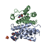 7vrzC 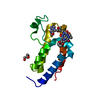 7vs0C 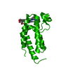 7vs1C 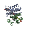 7vsfC 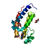 5xhkS S: Starting model for refinement C: citing same article ( |
|---|---|
| Similar structure data |
-Links
 PDBj
PDBj
- Assembly
| Deposited unit | 
| |||||||||
|---|---|---|---|---|---|---|---|---|---|---|
| 1 | 
| |||||||||
| Unit cell |
| |||||||||
| Components on special symmetry positions |
|
-Components
| #1: Protein | Mass: 13524.474 Da / Num. of mol.: 1 Source method: isolated from a genetically manipulated source Source: (gene. exp.) Homo sapiens (human) / Gene: BRD2 / Plasmid: pET28a / Production host:  | ||||
|---|---|---|---|---|---|
| #2: Chemical |   Mass: 180.164 Da / Num. of mol.: 2 / Source method: obtained synthetically / Formula: C7H8N4O2 / Feature type: SUBJECT OF INVESTIGATION Mass: 180.164 Da / Num. of mol.: 2 / Source method: obtained synthetically / Formula: C7H8N4O2 / Feature type: SUBJECT OF INVESTIGATION#3: Water | ChemComp-HOH / |  Mass: 18.015 Da / Num. of mol.: 225 / Source method: isolated from a natural source / Formula: H2O Mass: 18.015 Da / Num. of mol.: 225 / Source method: isolated from a natural source / Formula: H2OHas ligand of interest | Y | |
-Experimental details
-Experiment
| Experiment | Method: X-RAY DIFFRACTION / Number of used crystals: 1 |
|---|
- Sample preparation
| Crystal | Density Matthews: 2.24 Å3/Da / Density % sol: 45.12 % |
|---|---|
| Crystal grow | Temperature: 300 K / Method: vapor diffusion, hanging drop / Details: PEG MME 2000, 50mM Tris, 50mM NaCl |
-Data collection
| Diffraction | Mean temperature: 100 K / Serial crystal experiment: N |
|---|---|
| Diffraction source | Source: ROTATING ANODE / Type: RIGAKU MICROMAX-007 HF / Wavelength: 1.54056 Å |
| Detector | Type: RIGAKU HyPix-6000HE / Detector: PIXEL / Date: Aug 28, 2020 |
| Radiation | Protocol: SINGLE WAVELENGTH / Monochromatic (M) / Laue (L): M / Scattering type: x-ray |
| Radiation wavelength | Wavelength: 1.54056 Å / Relative weight: 1 |
| Reflection | Resolution: 1.1→25.61 Å / Num. obs: 48535 / % possible obs: 96 % / Redundancy: 3.6 % / CC1/2: 1 / Rmerge(I) obs: 0.068 / Net I/σ(I): 23.11 |
| Reflection shell | Resolution: 1.1→1.12 Å / Redundancy: 2.3 % / Rmerge(I) obs: 0.339 / Num. unique obs: 2015 / CC1/2: 0.95 / % possible all: 81 |
- Processing
| Software |
| |||||||||||||||||||||||||||||||||||||||||||||||||||||||||||||||||||||||||||||||||||||||||||||||||||||||||
|---|---|---|---|---|---|---|---|---|---|---|---|---|---|---|---|---|---|---|---|---|---|---|---|---|---|---|---|---|---|---|---|---|---|---|---|---|---|---|---|---|---|---|---|---|---|---|---|---|---|---|---|---|---|---|---|---|---|---|---|---|---|---|---|---|---|---|---|---|---|---|---|---|---|---|---|---|---|---|---|---|---|---|---|---|---|---|---|---|---|---|---|---|---|---|---|---|---|---|---|---|---|---|---|---|---|---|
| Refinement | Method to determine structure: MOLECULAR REPLACEMENT Starting model: 5XHK Resolution: 1.1→25.61 Å / SU ML: 0.08 / Cross valid method: FREE R-VALUE / σ(F): 1.34 / Phase error: 17.46 / Stereochemistry target values: ML
| |||||||||||||||||||||||||||||||||||||||||||||||||||||||||||||||||||||||||||||||||||||||||||||||||||||||||
| Solvent computation | Shrinkage radii: 0.9 Å / VDW probe radii: 1.11 Å / Solvent model: FLAT BULK SOLVENT MODEL | |||||||||||||||||||||||||||||||||||||||||||||||||||||||||||||||||||||||||||||||||||||||||||||||||||||||||
| Refinement step | Cycle: LAST / Resolution: 1.1→25.61 Å
| |||||||||||||||||||||||||||||||||||||||||||||||||||||||||||||||||||||||||||||||||||||||||||||||||||||||||
| Refine LS restraints |
| |||||||||||||||||||||||||||||||||||||||||||||||||||||||||||||||||||||||||||||||||||||||||||||||||||||||||
| LS refinement shell |
|
