 Movie
Movie Controller

[English] 日本語
 Yorodumi
- PDB-7vr6: Crystal structure of MlaC from Escherichia coli in quasi-open state -
+ Open data

- Basic information
| Entry | Database: PDB / ID: 7vr6 | ||||||
|---|---|---|---|---|---|---|---|
| Title | Crystal structure of MlaC from Escherichia coli in quasi-open state | ||||||
 Components Components | Intermembrane phospholipid transport system binding protein MlaC | ||||||
 Keywords Keywords | TRANSPORT PROTEIN / ABC transporter / Periplasmic protein / Membrane lipid asymmetry / Segmented domain movement / Mla transport system | ||||||
| Function / homology | Tgt2/MlaC superfamily / Toluene tolerance Ttg2/phospholipid-binding protein MlaC / MlaC protein / intermembrane phospholipid transfer / phospholipid transport / outer membrane-bounded periplasmic space / DI-PALMITOYL-3-SN-PHOSPHATIDYLETHANOLAMINE / Intermembrane phospholipid transport system binding protein MlaC Function and homology information Function and homology information | ||||||
| Biological species |  | ||||||
| Method |  X-RAY DIFFRACTION / MOLECULAR REPLACEMENT / molecular replacement / Resolution: 2.5 Å X-RAY DIFFRACTION / MOLECULAR REPLACEMENT / molecular replacement / Resolution: 2.5 Å | ||||||
 Authors Authors | Dutta, A. / Kanaujia, S.P. | ||||||
| Funding support |  India, 1items India, 1items
| ||||||
 Citation Citation | Journal: J.Struct.Biol. / Year: 2022 Title: MlaC belongs to a unique class of non-canonical substrate-binding proteins and follows a novel phospholipid-binding mechanism. Authors: Dutta, A. / Prasad Kanaujia, S. | ||||||
| History |
|
- Structure visualization
| Structure viewer | Molecule: MolmilJmol/JSmol |
|---|
- Downloads & links
-Download
| PDBx/mmCIF format | 7vr6.cif.gz | 90 KB | Display | PDBx/mmCIF format |
|---|---|---|---|---|
| PDB format | pdb7vr6.ent.gz | 65.4 KB | Display | PDB format |
| PDBx/mmJSON format | 7vr6.json.gz | Tree view | PDBx/mmJSON format | |
| Others |  Other downloads Other downloads |
-Validation report
| Arichive directory | https://data.pdbj.org/pub/pdb/validation_reports/vr/7vr6ftp://data.pdbj.org/pub/pdb/validation_reports/vr/7vr6 | HTTPS FTP |
|---|
-Related structure data
| Related structure data | 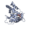 5uwaS S: Starting model for refinement |
|---|---|
| Similar structure data |
-Links
 PDBj
PDBj- Assembly
| Deposited unit | 
| ||||||||
|---|---|---|---|---|---|---|---|---|---|
| 1 |
| ||||||||
| Unit cell |
|
-Components
| #1: Protein | Mass: 22717.760 Da / Num. of mol.: 1 Source method: isolated from a genetically manipulated source Source: (gene. exp.) |
|---|---|
| #2: Chemical | ChemComp-PEF /   Mass: 691.959 Da / Num. of mol.: 1 / Source method: obtained synthetically / Formula: C37H74NO8P / Feature type: SUBJECT OF INVESTIGATION / Comment: phospholipid*YM Mass: 691.959 Da / Num. of mol.: 1 / Source method: obtained synthetically / Formula: C37H74NO8P / Feature type: SUBJECT OF INVESTIGATION / Comment: phospholipid*YM |
| #3: Chemical | ChemComp-EDO /   Mass: 62.068 Da / Num. of mol.: 1 / Source method: obtained synthetically / Formula: C2H6O2 Mass: 62.068 Da / Num. of mol.: 1 / Source method: obtained synthetically / Formula: C2H6O2 |
| #4: Water | ChemComp-HOH /  Mass: 18.015 Da / Num. of mol.: 58 / Source method: isolated from a natural source / Formula: H2O Mass: 18.015 Da / Num. of mol.: 58 / Source method: isolated from a natural source / Formula: H2O |
| Has ligand of interest | Y |
-Experimental details
-Experiment
| Experiment | Method: X-RAY DIFFRACTION / Number of used crystals: 1 |
|---|
- Sample preparation
| Crystal | Density Matthews: 2.72 Å3/Da / Density % sol: 54.7 % Description: THE ENTRY CONTAINS FRIEDEL PAIRS IN I/F_PLUS/MINUS COLUMNS. |
|---|---|
| Crystal grow | Temperature: 293 K / Method: vapor diffusion, hanging drop / pH: 7 Details: 0.7 M sodium citrate tribasic dihydrate, 0.1 M Bis-Tris propane pH 7.0 |
-Data collection
| Diffraction | Mean temperature: 100 K / Serial crystal experiment: N | |||||||||||||||||||||||||||
|---|---|---|---|---|---|---|---|---|---|---|---|---|---|---|---|---|---|---|---|---|---|---|---|---|---|---|---|---|
| Diffraction source | Source: ROTATING ANODE / Type: RIGAKU MICROMAX-007 HF / Wavelength: 1.5418 Å | |||||||||||||||||||||||||||
| Detector | Type: RIGAKU RAXIS IV++ / Detector: IMAGE PLATE / Date: Mar 28, 2018 / Details: VariMax HF | |||||||||||||||||||||||||||
| Radiation | Monochromator: Ni filter / Protocol: SINGLE WAVELENGTH / Monochromatic (M) / Laue (L): M / Scattering type: x-ray | |||||||||||||||||||||||||||
| Radiation wavelength | Wavelength: 1.5418 Å / Relative weight: 1 | |||||||||||||||||||||||||||
| Reflection | Resolution: 2.5→57.47 Å / Num. obs: 7857 / % possible obs: 100 % / Redundancy: 4.5 % / CC1/2: 0.996 / Rmerge(I) obs: 0.095 / Rpim(I) all: 0.05 / Rrim(I) all: 0.108 / Net I/σ(I): 13.5 / Num. measured all: 35454 / Scaling rejects: 26 | |||||||||||||||||||||||||||
| Reflection shell | Diffraction-ID: 1 / Redundancy: 4.5 %
|
-Phasing
| Phasing | Method: molecular replacement | |||||||||
|---|---|---|---|---|---|---|---|---|---|---|
| Phasing MR | Model details: Phaser MODE: MR_AUTO
|
- Processing
| Software |
| ||||||||||||||||||||||||||||||||||||||||||||||||||||||||||||
|---|---|---|---|---|---|---|---|---|---|---|---|---|---|---|---|---|---|---|---|---|---|---|---|---|---|---|---|---|---|---|---|---|---|---|---|---|---|---|---|---|---|---|---|---|---|---|---|---|---|---|---|---|---|---|---|---|---|---|---|---|---|
| Refinement | Method to determine structure: MOLECULAR REPLACEMENT Starting model: 5UWA Resolution: 2.5→57.47 Å / Cor.coef. Fo:Fc: 0.952 / Cor.coef. Fo:Fc free: 0.916 / SU B: 16.892 / SU ML: 0.186 / SU R Cruickshank DPI: 0.54 / Cross valid method: THROUGHOUT / σ(F): 0 / ESU R: 0.54 / ESU R Free: 0.256 / Stereochemistry target values: MAXIMUM LIKELIHOOD Details: U VALUES : WITH TLS ADDED HYDROGENS HAVE BEEN ADDED IN THE RIDING POSITIONS
| ||||||||||||||||||||||||||||||||||||||||||||||||||||||||||||
| Solvent computation | Ion probe radii: 0.8 Å / Shrinkage radii: 0.8 Å / VDW probe radii: 1.2 Å / Solvent model: MASK | ||||||||||||||||||||||||||||||||||||||||||||||||||||||||||||
| Displacement parameters | Biso max: 96.04 Å2 / Biso mean: 31.629 Å2 / Biso min: 13.3 Å2
| ||||||||||||||||||||||||||||||||||||||||||||||||||||||||||||
| Refinement step | Cycle: final / Resolution: 2.5→57.47 Å
| ||||||||||||||||||||||||||||||||||||||||||||||||||||||||||||
| Refine LS restraints |
| ||||||||||||||||||||||||||||||||||||||||||||||||||||||||||||
| LS refinement shell | Resolution: 2.501→2.565 Å / Rfactor Rfree error: 0 / Total num. of bins used: 20
| ||||||||||||||||||||||||||||||||||||||||||||||||||||||||||||
| Refinement TLS params. | Method: refined / Origin x: 6.431 Å / Origin y: 19.088 Å / Origin z: 4.362 Å
|
