 Movie
Movie Controller

[English] 日本語
 Yorodumi
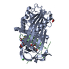
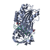
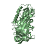
- PDB-7vlj: Crystal structure of Entamoeba histolytica serine protease inhibi... -
+ Open data

- Basic information
| Entry | Database: PDB / ID: 7vlj | |||||||||
|---|---|---|---|---|---|---|---|---|---|---|
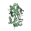
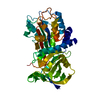
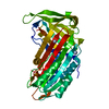
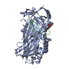
| Title | Crystal structure of Entamoeba histolytica serine protease inhibitor, Histopin, in the cleaved conformation | |||||||||
 Components Components | (Serine protease inhibitor, ...) x 2 | |||||||||
 Keywords Keywords | HYDROLASE / Serine protease inhibitor / Serine endopeptidases / Peptidase inhibitors / Serpin family I4 | |||||||||
| Function / homology |  Function and homology information Function and homology information | |||||||||
| Biological species |   Entamoeba histolytica (eukaryote) Entamoeba histolytica (eukaryote) | |||||||||
| Method |  X-RAY DIFFRACTION / SYNCHROTRON / MOLECULAR REPLACEMENT / Resolution: 1.83 Å X-RAY DIFFRACTION / SYNCHROTRON / MOLECULAR REPLACEMENT / Resolution: 1.83 Å | |||||||||
 Authors Authors | Ali, M.F. / Devi, S. / Gourinath, S. | |||||||||
| Funding support |  India, 1items India, 1items
| |||||||||
 Citation Citation | Journal: To Be Published Title: Crystal structure of Entamoeba histolytica serine protease inhibitor, Histopin, in the cleaved conformation Authors: Ali, M.F. / Devi, S. / Gourinath, S. | |||||||||
| History |
|
- Structure visualization
| Structure viewer | Molecule: MolmilJmol/JSmol |
|---|
- Downloads & links
-Download
| PDBx/mmCIF format | 7vlj.cif.gz | 94.9 KB | Display | PDBx/mmCIF format |
|---|---|---|---|---|
| PDB format | pdb7vlj.ent.gz | 68.9 KB | Display | PDB format |
| PDBx/mmJSON format | 7vlj.json.gz | Tree view | PDBx/mmJSON format | |
| Others |  Other downloads Other downloads |
-Validation report
| Arichive directory | https://data.pdbj.org/pub/pdb/validation_reports/vl/7vljftp://data.pdbj.org/pub/pdb/validation_reports/vl/7vlj | HTTPS FTP |
|---|
-Related structure data
| Related structure data | 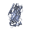 3ndaS S: Starting model for refinement |
|---|---|
| Similar structure data |


-Links
 PDBj
PDBj

- Assembly
| Deposited unit | 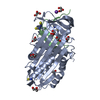
| ||||||||
|---|---|---|---|---|---|---|---|---|---|
| 1 |
| ||||||||
| Unit cell |
|
-Components
-Serine protease inhibitor, ... , 2 types, 2 molecules AB
| #1: Protein | Mass: 37760.359 Da / Num. of mol.: 1 Source method: isolated from a genetically manipulated source Source: (gene. exp.) Entamoeba histolytica (eukaryote) / Gene: EHI_119330 / Production host:  |
|---|---|
| #2: Protein/peptide | Mass: 5972.965 Da / Num. of mol.: 1 Source method: isolated from a genetically manipulated source Source: (gene. exp.) Entamoeba histolytica (eukaryote) / Gene: EHI_119330 / Production host: |
-Non-polymers , 5 types, 233 molecules 


| #3: Chemical | ChemComp-K /  Mass: 39.098 Da / Num. of mol.: 1 / Source method: obtained synthetically / Formula: K Mass: 39.098 Da / Num. of mol.: 1 / Source method: obtained synthetically / Formula: K | ||||||
|---|---|---|---|---|---|---|---|
| #4: Chemical | ChemComp-GOL /  Mass: 92.094 Da / Num. of mol.: 6 / Source method: obtained synthetically / Formula: C3H8O3 Mass: 92.094 Da / Num. of mol.: 6 / Source method: obtained synthetically / Formula: C3H8O3#5: Chemical |  Mass: 106.120 Da / Num. of mol.: 3 / Source method: obtained synthetically / Formula: C4H10O3 Mass: 106.120 Da / Num. of mol.: 3 / Source method: obtained synthetically / Formula: C4H10O3#6: Chemical | ChemComp-SCN /  Mass: 58.082 Da / Num. of mol.: 5 / Source method: obtained synthetically / Formula: CNS Mass: 58.082 Da / Num. of mol.: 5 / Source method: obtained synthetically / Formula: CNS#7: Water | ChemComp-HOH / | Mass: 18.015 Da / Num. of mol.: 218 / Source method: isolated from a natural source / Formula: H2O |
-Details
| Has ligand of interest | N |
|---|
-Experimental details
-Experiment
| Experiment | Method: X-RAY DIFFRACTION / Number of used crystals: 1 |
|---|
- Sample preparation
| Crystal | Density Matthews: 1.96 Å3/Da / Density % sol: 37.37 % |
|---|---|
| Crystal grow | Temperature: 289 K / Method: vapor diffusion, hanging drop Details: 25% Polyethylene glycol monomethyl ether 2000, 0.2M Potassium thiocyanate |
-Data collection
| Diffraction | Mean temperature: 100 K / Serial crystal experiment: N |
|---|---|
| Diffraction source | Source: SYNCHROTRON / Site: ESRF  / Beamline: MASSIF-3 / Wavelength: 0.9677 Å / Beamline: MASSIF-3 / Wavelength: 0.9677 Å |
| Detector | Type: DECTRIS EIGER X 4M / Detector: PIXEL / Date: Apr 7, 2018 |
| Radiation | Monochromator: single Silicon (111) / Protocol: SINGLE WAVELENGTH / Monochromatic (M) / Laue (L): M / Scattering type: x-ray |
| Radiation wavelength | Wavelength: 0.9677 Å / Relative weight: 1 |
| Reflection | Resolution: 1.83→45.64 Å / Num. obs: 29415 / % possible obs: 98.5 % / Redundancy: 7.3 % / CC1/2: 0.998 / Rmerge(I) obs: 0.066 / Net I/σ(I): 17.2 |
| Reflection shell | Resolution: 1.83→1.87 Å / Redundancy: 7.1 % / Num. unique obs: 1673 / CC1/2: 0.977 / % possible all: 92 |
- Processing
| Software |
| ||||||||||||||||||||||||||||||||||||||||||||||||||||||||||||
|---|---|---|---|---|---|---|---|---|---|---|---|---|---|---|---|---|---|---|---|---|---|---|---|---|---|---|---|---|---|---|---|---|---|---|---|---|---|---|---|---|---|---|---|---|---|---|---|---|---|---|---|---|---|---|---|---|---|---|---|---|---|
| Refinement | Method to determine structure: MOLECULAR REPLACEMENT Starting model: 3NDA Resolution: 1.83→45.64 Å / Cor.coef. Fo:Fc: 0.961 / Cor.coef. Fo:Fc free: 0.943 / SU B: 2.411 / SU ML: 0.075 / Cross valid method: THROUGHOUT / σ(F): 0 / ESU R: 0.137 / ESU R Free: 0.129 / Stereochemistry target values: MAXIMUM LIKELIHOOD Details: HYDROGENS HAVE BEEN ADDED IN THE RIDING POSITIONS U VALUES : REFINED INDIVIDUALLY
| ||||||||||||||||||||||||||||||||||||||||||||||||||||||||||||
| Solvent computation | Ion probe radii: 0.8 Å / Shrinkage radii: 0.8 Å / VDW probe radii: 1.2 Å / Solvent model: MASK | ||||||||||||||||||||||||||||||||||||||||||||||||||||||||||||
| Displacement parameters | Biso max: 75.13 Å2 / Biso mean: 18.274 Å2 / Biso min: 7.89 Å2
| ||||||||||||||||||||||||||||||||||||||||||||||||||||||||||||
| Refinement step | Cycle: final / Resolution: 1.83→45.64 Å
| ||||||||||||||||||||||||||||||||||||||||||||||||||||||||||||
| Refine LS restraints |
| ||||||||||||||||||||||||||||||||||||||||||||||||||||||||||||
| LS refinement shell | Resolution: 1.83→1.878 Å / Rfactor Rfree error: 0 / Total num. of bins used: 20
|
