 Movie
Movie Controller

[English] 日本語
 Yorodumi
- PDB-7vgv: Anion free form of light-driven chloride ion-pumping rhodopsin, N... -
+ Open data

- Basic information
| Entry | Database: PDB / ID: 7vgv | ||||||
|---|---|---|---|---|---|---|---|
| Title | Anion free form of light-driven chloride ion-pumping rhodopsin, NM-R3, structure determined by serial femtosecond crystallography at SACLA | ||||||
 Components Components | Chloride pumping rhodopsin | ||||||
 Keywords Keywords | MEMBRANE PROTEIN / SACLA serial femtosecond crystallography CELL-FREE SYNTHESIS Bacterial type rhodopsin chloride ion pump rhodopsin | ||||||
| Function / homology |  Function and homology information Function and homology information | ||||||
| Biological species |  Nonlabens marinus S1-08 (bacteria) Nonlabens marinus S1-08 (bacteria) | ||||||
| Method |  X-RAY DIFFRACTION / FREE ELECTRON LASER / MOLECULAR REPLACEMENT / Resolution: 2.3 Å X-RAY DIFFRACTION / FREE ELECTRON LASER / MOLECULAR REPLACEMENT / Resolution: 2.3 Å | ||||||
 Authors Authors | Hosaka, T. / Nango, E. / Nakane, T. / Luo, F. / Kimura-Someya, T. / Shirouzu, M. | ||||||
| Funding support | 1items
| ||||||
 Citation Citation | Journal: Proc.Natl.Acad.Sci.USA / Year: 2022 Title: Conformational alterations in unidirectional ion transport of a light-driven chloride pump revealed using X-ray free electron lasers. Authors: Hosaka, T. / Nomura, T. / Kubo, M. / Nakane, T. / Fangjia, L. / Sekine, S.I. / Ito, T. / Murayama, K. / Ihara, K. / Ehara, H. / Kashiwagi, K. / Katsura, K. / Akasaka, R. / Hisano, T. / ...Authors: Hosaka, T. / Nomura, T. / Kubo, M. / Nakane, T. / Fangjia, L. / Sekine, S.I. / Ito, T. / Murayama, K. / Ihara, K. / Ehara, H. / Kashiwagi, K. / Katsura, K. / Akasaka, R. / Hisano, T. / Tanaka, T. / Tanaka, R. / Arima, T. / Yamashita, A. / Sugahara, M. / Naitow, H. / Matsuura, Y. / Yoshizawa, S. / Tono, K. / Owada, S. / Nureki, O. / Kimura-Someya, T. / Iwata, S. / Nango, E. / Shirouzu, M. | ||||||
| History |
|
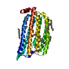
- Structure visualization
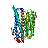
| Structure viewer | Molecule: MolmilJmol/JSmol |
|---|
- Downloads & links
-Download
| PDBx/mmCIF format | 7vgv.cif.gz | 171.6 KB | Display | PDBx/mmCIF format |
|---|---|---|---|---|
| PDB format | pdb7vgv.ent.gz | 133.6 KB | Display | PDB format |
| PDBx/mmJSON format | 7vgv.json.gz | Tree view | PDBx/mmJSON format | |
| Others |  Other downloads Other downloads |
-Validation report
| Arichive directory | https://data.pdbj.org/pub/pdb/validation_reports/vg/7vgvftp://data.pdbj.org/pub/pdb/validation_reports/vg/7vgv | HTTPS FTP |
|---|
-Related structure data
| Related structure data |  7vgtC  7vguC 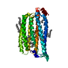 5b2nS S: Starting model for refinement C: citing same article ( |
|---|---|
| Similar structure data | |
| Experimental dataset #1 | Data reference: 10.11577/1843782 / Data set type: diffraction image data |

-Links
 PDBj
PDBj


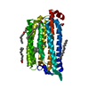
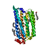
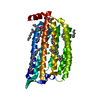
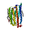
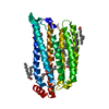
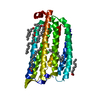
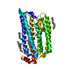
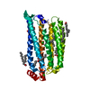
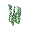
- Assembly
| Deposited unit | 
| ||||||||
|---|---|---|---|---|---|---|---|---|---|
| 1 | 
| ||||||||
| 2 | 
| ||||||||
| 3 | 
| ||||||||
| Unit cell |
|
-Components
-Protein , 1 types, 3 molecules ABC
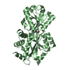
| #1: Protein | Mass: 30710.936 Da / Num. of mol.: 3 Source method: isolated from a genetically manipulated source Source: (gene. exp.) Nonlabens marinus S1-08 (bacteria) / Gene: ClR, NMS_1267 / Production host: |
|---|
-Non-polymers , 8 types, 88 molecules 
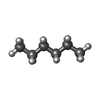
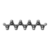


| #2: Chemical |  Mass: 284.436 Da / Num. of mol.: 3 / Source method: obtained synthetically / Formula: C20H28O Mass: 284.436 Da / Num. of mol.: 3 / Source method: obtained synthetically / Formula: C20H28O#3: Chemical | ChemComp-HEX /  Mass: 86.175 Da / Num. of mol.: 6 / Source method: obtained synthetically / Formula: C6H14 Mass: 86.175 Da / Num. of mol.: 6 / Source method: obtained synthetically / Formula: C6H14#4: Chemical |  Mass: 128.255 Da / Num. of mol.: 3 / Source method: obtained synthetically / Formula: C9H20 Mass: 128.255 Da / Num. of mol.: 3 / Source method: obtained synthetically / Formula: C9H20#5: Chemical |  Mass: 198.388 Da / Num. of mol.: 2 / Source method: obtained synthetically / Formula: C14H30 Mass: 198.388 Da / Num. of mol.: 2 / Source method: obtained synthetically / Formula: C14H30#6: Chemical | ChemComp-R16 / |  Mass: 226.441 Da / Num. of mol.: 1 / Source method: obtained synthetically / Formula: C16H34 Mass: 226.441 Da / Num. of mol.: 1 / Source method: obtained synthetically / Formula: C16H34#7: Chemical | ChemComp-OCT / |  Mass: 114.229 Da / Num. of mol.: 1 / Source method: obtained synthetically / Formula: C8H18 Mass: 114.229 Da / Num. of mol.: 1 / Source method: obtained synthetically / Formula: C8H18#8: Chemical |  Mass: 35.453 Da / Num. of mol.: 2 / Source method: obtained synthetically / Formula: Cl Mass: 35.453 Da / Num. of mol.: 2 / Source method: obtained synthetically / Formula: Cl#9: Water | ChemComp-HOH / | Mass: 18.015 Da / Num. of mol.: 70 / Source method: isolated from a natural source / Formula: H2O |
|---|
-Details
| Has ligand of interest | N |
|---|---|
| Has protein modification | Y |
-Experimental details
-Experiment
| Experiment | Method: X-RAY DIFFRACTION / Number of used crystals: 1 |
|---|
- Sample preparation
| Crystal | Density Matthews: 2.98 Å3/Da / Density % sol: 58.79 % |
|---|---|
| Crystal grow | Temperature: 293 K / Method: lipidic cubic phase Details: 100 mM HEPES (pH7.0), 400-700 mM K phosphate, and 24-30% PEG400 |
-Data collection
| Diffraction | Mean temperature: 293 K / Serial crystal experiment: Y |
|---|---|
| Diffraction source | Source: FREE ELECTRON LASER / Site: SACLA  / Beamline: BL3 / Wavelength: 1.771 Å / Beamline: BL3 / Wavelength: 1.771 Å |
| Detector | Type: MPCCD / Detector: CCD / Date: May 10, 2016 |
| Radiation | Protocol: SINGLE WAVELENGTH / Monochromatic (M) / Laue (L): M / Scattering type: x-ray |
| Radiation wavelength | Wavelength: 1.771 Å / Relative weight: 1 |
| Reflection | Resolution: 2.3→46.3 Å / Num. obs: 50102 / % possible obs: 100 % / Redundancy: 136 % / CC1/2: 0.984 / Net I/σ(I): 5.41 |
| Reflection shell | Resolution: 2.3→2.34 Å / Num. unique obs: 2805 / CC1/2: 0.473 |
| Serial crystallography sample delivery | Method: injection |
- Processing
| Software |
| |||||||||||||||||||||||||||||||||||||||||||||||||||||||||||||||||||||||||||||||||||||||||||||||||||||||||||||||||||||||||||||||||||||
|---|---|---|---|---|---|---|---|---|---|---|---|---|---|---|---|---|---|---|---|---|---|---|---|---|---|---|---|---|---|---|---|---|---|---|---|---|---|---|---|---|---|---|---|---|---|---|---|---|---|---|---|---|---|---|---|---|---|---|---|---|---|---|---|---|---|---|---|---|---|---|---|---|---|---|---|---|---|---|---|---|---|---|---|---|---|---|---|---|---|---|---|---|---|---|---|---|---|---|---|---|---|---|---|---|---|---|---|---|---|---|---|---|---|---|---|---|---|---|---|---|---|---|---|---|---|---|---|---|---|---|---|---|---|---|
| Refinement | Method to determine structure: MOLECULAR REPLACEMENT Starting model: 5B2N Resolution: 2.3→44.46 Å / SU ML: 0.26 / Cross valid method: THROUGHOUT / σ(F): 1.97 / Phase error: 24.72 / Stereochemistry target values: ML
| |||||||||||||||||||||||||||||||||||||||||||||||||||||||||||||||||||||||||||||||||||||||||||||||||||||||||||||||||||||||||||||||||||||
| Solvent computation | Shrinkage radii: 0.9 Å / VDW probe radii: 1.11 Å / Solvent model: FLAT BULK SOLVENT MODEL | |||||||||||||||||||||||||||||||||||||||||||||||||||||||||||||||||||||||||||||||||||||||||||||||||||||||||||||||||||||||||||||||||||||
| Displacement parameters | Biso max: 127.62 Å2 / Biso mean: 53.833 Å2 / Biso min: 26.77 Å2 | |||||||||||||||||||||||||||||||||||||||||||||||||||||||||||||||||||||||||||||||||||||||||||||||||||||||||||||||||||||||||||||||||||||
| Refinement step | Cycle: final / Resolution: 2.3→44.46 Å
| |||||||||||||||||||||||||||||||||||||||||||||||||||||||||||||||||||||||||||||||||||||||||||||||||||||||||||||||||||||||||||||||||||||
| LS refinement shell | Refine-ID: X-RAY DIFFRACTION / Rfactor Rfree error: 0 / Total num. of bins used: 18
|
