 Movie
Movie Controller

[English] 日本語
 Yorodumi
- PDB-7vgc: Crystal structure of prolyl oligopeptidase from Microbulbifer are... -
+ Open data

- Basic information
| Entry | Database: PDB / ID: 7vgc | ||||||
|---|---|---|---|---|---|---|---|
| Title | Crystal structure of prolyl oligopeptidase from Microbulbifer arenaceous complex with a transition state analog inhibitor ZPR | ||||||
 Components Components | prolyl oligopeptidase | ||||||
 Keywords Keywords | HYDROLASE / S9A / prolyl endopeptidase / serine protease / mental disorder / amnesia | ||||||
| Function / homology | Z-PRO-PROLINAL / N-BENZYLOXYCARBONYL-L-PROLYL-L-PROLINAL Function and homology information Function and homology information | ||||||
| Biological species |  Microbulbifer arenaceous (bacteria) Microbulbifer arenaceous (bacteria) | ||||||
| Method |  X-RAY DIFFRACTION / SYNCHROTRON / MOLECULAR REPLACEMENT / Resolution: 2.722 Å X-RAY DIFFRACTION / SYNCHROTRON / MOLECULAR REPLACEMENT / Resolution: 2.722 Å | ||||||
 Authors Authors | Huang, P. / Yang, S.Q. / Jiang, Z.Q. | ||||||
| Funding support |  China, 1items China, 1items
| ||||||
 Citation Citation | Journal: Acta Crystallogr D Struct Biol / Year: 2022 Title: The structure and molecular dynamics of prolyl oligopeptidase from Microbulbifer arenaceous provide insights into catalytic and regulatory mechanisms. Authors: Huang, P. / Lv, A. / Yan, Q. / Jiang, Z. / Yang, S. | ||||||
| History |
|
- Structure visualization
| Structure viewer | Molecule: MolmilJmol/JSmol |
|---|
- Downloads & links
-Download
| PDBx/mmCIF format | 7vgc.cif.gz | 154.2 KB | Display | PDBx/mmCIF format |
|---|---|---|---|---|
| PDB format | pdb7vgc.ent.gz | 114.5 KB | Display | PDB format |
| PDBx/mmJSON format | 7vgc.json.gz | Tree view | PDBx/mmJSON format | |
| Others |  Other downloads Other downloads |
-Validation report
| Arichive directory | https://data.pdbj.org/pub/pdb/validation_reports/vg/7vgcftp://data.pdbj.org/pub/pdb/validation_reports/vg/7vgc | HTTPS FTP |
|---|
-Related structure data
| Related structure data | 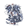 7vgbSC S: Starting model for refinement C: citing same article ( |
|---|---|
| Similar structure data |
-Links
 PDBj
PDBj- Assembly
| Deposited unit | 
| ||||||||
|---|---|---|---|---|---|---|---|---|---|
| 1 |
| ||||||||
| Unit cell |
|
-Components
| #1: Protein | Mass: 79952.180 Da / Num. of mol.: 1 Source method: isolated from a genetically manipulated source Source: (gene. exp.) Microbulbifer arenaceous (bacteria)Production host: References: prolyl oligopeptidase | ||||||
|---|---|---|---|---|---|---|---|
| #2: Chemical | ChemComp-ZPR / 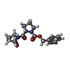  Type: peptide-like, Peptide-like / Class: Inhibitor / Mass: 330.378 Da / Num. of mol.: 1 / Source method: obtained synthetically / Formula: C18H22N2O4 / Feature type: SUBJECT OF INVESTIGATION / References: Z-PRO-PROLINAL Type: peptide-like, Peptide-like / Class: Inhibitor / Mass: 330.378 Da / Num. of mol.: 1 / Source method: obtained synthetically / Formula: C18H22N2O4 / Feature type: SUBJECT OF INVESTIGATION / References: Z-PRO-PROLINAL | ||||||
| #3: Chemical |   Mass: 35.453 Da / Num. of mol.: 2 / Source method: obtained synthetically / Formula: Cl Mass: 35.453 Da / Num. of mol.: 2 / Source method: obtained synthetically / Formula: Cl#4: Water | ChemComp-HOH / |  Mass: 18.015 Da / Num. of mol.: 67 / Source method: isolated from a natural source / Formula: H2O Mass: 18.015 Da / Num. of mol.: 67 / Source method: isolated from a natural source / Formula: H2OHas ligand of interest | Y | Has protein modification | Y | |
-Experimental details
-Experiment
| Experiment | Method: X-RAY DIFFRACTION / Number of used crystals: 1 |
|---|
- Sample preparation
| Crystal | Density Matthews: 2.26 Å3/Da / Density % sol: 45.48 % |
|---|---|
| Crystal grow | Temperature: 277 K / Method: vapor diffusion, hanging drop Details: 0.04M MES monohydrate pH 6.0, 8.8% (v/v) Polyethylene glycol 400 |
-Data collection
| Diffraction | Mean temperature: 100 K / Serial crystal experiment: N |
|---|---|
| Diffraction source | Source: SYNCHROTRON / Site: SSRF / Beamline: BL17B1 / Wavelength: 0.979 Å |
| Detector | Type: DECTRIS PILATUS3 S 2M / Detector: PIXEL / Date: Jun 27, 2021 |
| Radiation | Protocol: SINGLE WAVELENGTH / Monochromatic (M) / Laue (L): M / Scattering type: x-ray |
| Radiation wavelength | Wavelength: 0.979 Å / Relative weight: 1 |
| Reflection | Resolution: 2.722→30.96 Å / Num. obs: 18620 / % possible obs: 96.28 % / Redundancy: 5.1 % / CC1/2: 0.924 / CC star: 0.98 / Net I/σ(I): 5.75 |
| Reflection shell | Resolution: 2.722→2.82 Å / Num. unique obs: 6850 / CC1/2: 0.924 |
- Processing
| Software |
| |||||||||||||||||||||||||||||||||||||||||||||||||||||||||||||||||||||||||||||||||||||||||||||||||||||||||||||||||||||||||||||||||||||||||||||||||||
|---|---|---|---|---|---|---|---|---|---|---|---|---|---|---|---|---|---|---|---|---|---|---|---|---|---|---|---|---|---|---|---|---|---|---|---|---|---|---|---|---|---|---|---|---|---|---|---|---|---|---|---|---|---|---|---|---|---|---|---|---|---|---|---|---|---|---|---|---|---|---|---|---|---|---|---|---|---|---|---|---|---|---|---|---|---|---|---|---|---|---|---|---|---|---|---|---|---|---|---|---|---|---|---|---|---|---|---|---|---|---|---|---|---|---|---|---|---|---|---|---|---|---|---|---|---|---|---|---|---|---|---|---|---|---|---|---|---|---|---|---|---|---|---|---|---|---|---|---|
| Refinement | Method to determine structure: MOLECULAR REPLACEMENT Starting model: 7VGB Resolution: 2.722→30.96 Å / Cor.coef. Fo:Fc: 0.934 / Cor.coef. Fo:Fc free: 0.873 / SU B: 18.113 / SU ML: 0.352 / Cross valid method: FREE R-VALUE / ESU R Free: 0.425 / Details: Hydrogens have not been used
| |||||||||||||||||||||||||||||||||||||||||||||||||||||||||||||||||||||||||||||||||||||||||||||||||||||||||||||||||||||||||||||||||||||||||||||||||||
| Solvent computation | Ion probe radii: 0.8 Å / Shrinkage radii: 0.8 Å / VDW probe radii: 1.2 Å / Solvent model: BABINET MODEL PLUS MASK | |||||||||||||||||||||||||||||||||||||||||||||||||||||||||||||||||||||||||||||||||||||||||||||||||||||||||||||||||||||||||||||||||||||||||||||||||||
| Displacement parameters | Biso mean: 44.608 Å2
| |||||||||||||||||||||||||||||||||||||||||||||||||||||||||||||||||||||||||||||||||||||||||||||||||||||||||||||||||||||||||||||||||||||||||||||||||||
| Refinement step | Cycle: LAST / Resolution: 2.722→30.96 Å
| |||||||||||||||||||||||||||||||||||||||||||||||||||||||||||||||||||||||||||||||||||||||||||||||||||||||||||||||||||||||||||||||||||||||||||||||||||
| Refine LS restraints |
| |||||||||||||||||||||||||||||||||||||||||||||||||||||||||||||||||||||||||||||||||||||||||||||||||||||||||||||||||||||||||||||||||||||||||||||||||||
| LS refinement shell |
|
