 Movie
Movie Controller

[English] 日本語
 Yorodumi
- PDB-7vg7: Plexin B1 extracellular fragment in complex with lasso-grafted PB... -
+ Open data

- Basic information
| Entry | Database: PDB / ID: 7vg7 | ||||||
|---|---|---|---|---|---|---|---|
| Title | Plexin B1 extracellular fragment in complex with lasso-grafted PB1m6A9 peptide | ||||||
 Components Components |
| ||||||
 Keywords Keywords | SIGNALING PROTEIN / Plexin / complex | ||||||
| Function / homology |  Function and homology information Function and homology informationpolychlorinated biphenyl binding / negative regulation of interleukin-4 production / negative regulation of interleukin-5 production / negative regulation of interleukin-13 production / phospholipase A2 inhibitor activity / semaphorin receptor binding / response to silicon dioxide / response to ozone / negative regulation of osteoblast proliferation / response to fibroblast growth factor ...polychlorinated biphenyl binding / negative regulation of interleukin-4 production / negative regulation of interleukin-5 production / negative regulation of interleukin-13 production / phospholipase A2 inhibitor activity / semaphorin receptor binding / response to silicon dioxide / response to ozone / negative regulation of osteoblast proliferation / response to fibroblast growth factor / semaphorin receptor complex / semaphorin receptor activity / negative regulation of cell adhesion / RHOD GTPase cycle / positive regulation of axonogenesis / Sema4D induced cell migration and growth-cone collapse / inhibitory synapse assembly / GTPase activating protein binding / Sema4D mediated inhibition of cell attachment and migration / ossification involved in bone maturation / regulation of cytoskeleton organization / positive regulation of Rho protein signal transduction / negative regulation of type II interferon production / semaphorin-plexin signaling pathway / positive regulation of GTPase activity / T cell proliferation / synapse assembly / negative regulation of T cell proliferation / neuron projection morphogenesis / response to cytokine / regulation of mRNA stability / embryo implantation / GTPase activator activity / regulation of cell migration / response to glucocorticoid / secretory granule / female pregnancy / transmembrane signaling receptor activity / nuclear envelope / cell migration / G alpha (12/13) signalling events / regulation of cell shape / regulation of inflammatory response / response to lipopolysaccharide / postsynaptic membrane / positive regulation of phosphatidylinositol 3-kinase/protein kinase B signal transduction / intracellular signal transduction / response to xenobiotic stimulus / glutamatergic synapse / negative regulation of transcription by RNA polymerase II / signal transduction / extracellular space / extracellular exosome / extracellular region / plasma membrane / cytoplasm Similarity search - Function | ||||||
| Biological species |  Homo sapiens (human) Homo sapiens (human) | ||||||
| Method |  X-RAY DIFFRACTION / SYNCHROTRON / MOLECULAR REPLACEMENT / Resolution: 2.5 Å X-RAY DIFFRACTION / SYNCHROTRON / MOLECULAR REPLACEMENT / Resolution: 2.5 Å | ||||||
 Authors Authors | Sugano, N.N. / Hirata, K. / Yamashita, K. / Yamamoto, M. / Arimori, T. / Takagi, J. | ||||||
| Funding support |  Japan, 1items Japan, 1items
| ||||||
 Citation Citation | Journal: Structure / Year: 2022 Title: De novo Fc-based receptor dimerizers differentially modulate PlexinB1 function. Authors: Sugano-Nakamura, N. / Matoba, K. / Hirose, M. / Bashiruddin, N.K. / Matsunaga, Y. / Yamashita, K. / Hirata, K. / Yamamoto, M. / Arimori, T. / Suga, H. / Takagi, J. | ||||||
| History |
|
- Structure visualization
| Structure viewer | Molecule: MolmilJmol/JSmol |
|---|
- Downloads & links
-Download
| PDBx/mmCIF format | 7vg7.cif.gz | 172.9 KB | Display | PDBx/mmCIF format |
|---|---|---|---|---|
| PDB format | pdb7vg7.ent.gz | 106.5 KB | Display | PDB format |
| PDBx/mmJSON format | 7vg7.json.gz | Tree view | PDBx/mmJSON format | |
| Others |  Other downloads Other downloads |
-Validation report
| Summary document | 7vg7_validation.pdf.gz | 470.3 KB | Display | wwPDB validaton report |
|---|---|---|---|---|
| Full document | 7vg7_full_validation.pdf.gz | 476.3 KB | Display | |
| Data in XML | 7vg7_validation.xml.gz | 25.3 KB | Display | |
| Data in CIF | 7vg7_validation.cif.gz | 34.9 KB | Display | |
| Arichive directory | https://data.pdbj.org/pub/pdb/validation_reports/vg/7vg7ftp://data.pdbj.org/pub/pdb/validation_reports/vg/7vg7 | HTTPS FTP |
-Related structure data
| Related structure data | 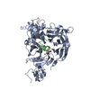 7vf3C  1utgS 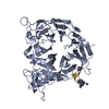 5b4wS C: citing same article ( S: Starting model for refinement |
|---|---|
| Similar structure data |
-Links
 PDBj
PDBj


- Assembly
| Deposited unit | 
| ||||||||||||
|---|---|---|---|---|---|---|---|---|---|---|---|---|---|
| 1 |
| ||||||||||||
| Unit cell |
|
-Components
| #1: Protein | Mass: 56661.395 Da / Num. of mol.: 1 / Mutation: T19S Source method: isolated from a genetically manipulated source Source: (gene. exp.) Homo sapiens (human) / Gene: PLXNB1, KIAA0407, PLXN5, SEP / Production host: Homo sapiens (human) / References: UniProt: O43157 |
|---|---|
| #2: Protein | Mass: 18300.041 Da / Num. of mol.: 1 / Mutation: A21S Source method: isolated from a genetically manipulated source Details: PB1m6A9-grafted uteroglobin / Source: (gene. exp.) Homo sapiens (human) / Gene: SCGB1A1, CC10, CCSP, UGB / Production host: Homo sapiens (human) / References: UniProt: P11684 |
| #3: Chemical | ChemComp-PGE /   Mass: 150.173 Da / Num. of mol.: 1 / Source method: obtained synthetically / Formula: C6H14O4 Mass: 150.173 Da / Num. of mol.: 1 / Source method: obtained synthetically / Formula: C6H14O4 |
| #4: Sugar | ChemComp-NAG /   Type: D-saccharide, beta linking / Mass: 221.208 Da / Num. of mol.: 1 / Source method: obtained synthetically / Formula: C8H15NO6 Type: D-saccharide, beta linking / Mass: 221.208 Da / Num. of mol.: 1 / Source method: obtained synthetically / Formula: C8H15NO6 |
| #5: Water | ChemComp-HOH /  Mass: 18.015 Da / Num. of mol.: 100 / Source method: isolated from a natural source / Formula: H2O Mass: 18.015 Da / Num. of mol.: 100 / Source method: isolated from a natural source / Formula: H2O |
| Has ligand of interest | N |
| Has protein modification | Y |
-Experimental details
-Experiment
| Experiment | Method: X-RAY DIFFRACTION / Number of used crystals: 1 |
|---|
- Sample preparation
| Crystal | Density Matthews: 2.21 Å3/Da / Density % sol: 44.34 % |
|---|---|
| Crystal grow | Temperature: 293 K / Method: vapor diffusion, hanging drop / Details: 0.1M MES or HEPES, 14-16% PEG 3350 / PH range: 6.5 - 7.0 |
-Data collection
| Diffraction | Mean temperature: 100 K / Serial crystal experiment: N |
|---|---|
| Diffraction source | Source: SYNCHROTRON / Site: SPring-8 / Beamline: BL32XU / Wavelength: 1 Å |
| Detector | Type: DECTRIS EIGER X 9M / Detector: PIXEL / Date: Feb 6, 2018 |
| Radiation | Protocol: SINGLE WAVELENGTH / Monochromatic (M) / Laue (L): M / Scattering type: x-ray |
| Radiation wavelength | Wavelength: 1 Å / Relative weight: 1 |
| Reflection | Resolution: 2.5→76.6 Å / Num. obs: 22866 / % possible obs: 99.8 % / Redundancy: 19.7 % / Biso Wilson estimate: 24.31 Å2 / CC1/2: 0.975 / Rrim(I) all: 0.473 / Net I/σ(I): 6.43 |
| Reflection shell | Resolution: 2.5→2.65 Å / Redundancy: 18.5 % / Num. unique obs: 3621 / CC1/2: 0.678 / % possible all: 99.8 |
- Processing
| Software |
| |||||||||||||||||||||||||||||||||||||||||||||||||||||||||||||||
|---|---|---|---|---|---|---|---|---|---|---|---|---|---|---|---|---|---|---|---|---|---|---|---|---|---|---|---|---|---|---|---|---|---|---|---|---|---|---|---|---|---|---|---|---|---|---|---|---|---|---|---|---|---|---|---|---|---|---|---|---|---|---|---|---|
| Refinement | Method to determine structure: MOLECULAR REPLACEMENT Starting model: 5B4W, 1UTG Resolution: 2.5→44.78 Å / SU ML: 0.3054 / Cross valid method: FREE R-VALUE / σ(F): 1.34 / Phase error: 24.0224 / Stereochemistry target values: GeoStd + Monomer Library
| |||||||||||||||||||||||||||||||||||||||||||||||||||||||||||||||
| Solvent computation | Shrinkage radii: 0.9 Å / VDW probe radii: 1.11 Å / Solvent model: FLAT BULK SOLVENT MODEL | |||||||||||||||||||||||||||||||||||||||||||||||||||||||||||||||
| Displacement parameters | Biso mean: 31.01 Å2 | |||||||||||||||||||||||||||||||||||||||||||||||||||||||||||||||
| Refinement step | Cycle: LAST / Resolution: 2.5→44.78 Å
| |||||||||||||||||||||||||||||||||||||||||||||||||||||||||||||||
| Refine LS restraints |
| |||||||||||||||||||||||||||||||||||||||||||||||||||||||||||||||
| LS refinement shell |
|
