 Movie
Movie Controller

[English] 日本語
 Yorodumi
- PDB-7vcr: Crystal Structure of PitA fragment from pilus islet-2 of Streptoc... -
+ Open data

- Basic information
| Entry | Database: PDB / ID: 7vcr | ||||||
|---|---|---|---|---|---|---|---|
| Title | Crystal Structure of PitA fragment from pilus islet-2 of Streptococcus oralis | ||||||
 Components Components | (von Willebrand factor type A domain protein) x 2 | ||||||
 Keywords Keywords | CELL ADHESION / Tip pilin / adhesin / PitA / PI-2 pilus / Streptococcus oralis / isopeptide / CnaA fold / CnaB fold / dental plaque / biofilm | ||||||
| Function / homology |  Function and homology information Function and homology information | ||||||
| Biological species |  Streptococcus oralis ATCC 35037 (bacteria) Streptococcus oralis ATCC 35037 (bacteria) | ||||||
| Method |  X-RAY DIFFRACTION / SYNCHROTRON / MOLECULAR REPLACEMENT / Resolution: 2 Å X-RAY DIFFRACTION / SYNCHROTRON / MOLECULAR REPLACEMENT / Resolution: 2 Å | ||||||
 Authors Authors | Yadav, R.K. / Krishnan, V. | ||||||
| Funding support |  India, 1items India, 1items
| ||||||
 Citation Citation | Journal: Febs J. / Year: 2022 Title: New structural insights into the PI-2 pilus from Streptococcus oralis, an early dental plaque colonizer. Authors: Yadav, R.K. / Krishnan, V. | ||||||
| History |
|
- Structure visualization
| Structure viewer | Molecule: MolmilJmol/JSmol |
|---|
- Downloads & links
-Download
| PDBx/mmCIF format | 7vcr.cif.gz | 368.4 KB | Display | PDBx/mmCIF format |
|---|---|---|---|---|
| PDB format | pdb7vcr.ent.gz | 291.8 KB | Display | PDB format |
| PDBx/mmJSON format | 7vcr.json.gz | Tree view | PDBx/mmJSON format | |
| Others |  Other downloads Other downloads |
-Validation report
| Arichive directory | https://data.pdbj.org/pub/pdb/validation_reports/vc/7vcrftp://data.pdbj.org/pub/pdb/validation_reports/vc/7vcr | HTTPS FTP |
|---|
-Related structure data
| Related structure data | 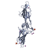 7f7yC 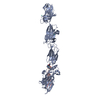 7vcnSC 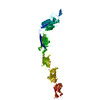 7w6bC  7w7iC S: Starting model for refinement C: citing same article ( |
|---|---|
| Similar structure data |
-Links
 PDBj
PDBj


- Assembly
| Deposited unit | 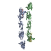
| ||||||||||||||||||||||||
|---|---|---|---|---|---|---|---|---|---|---|---|---|---|---|---|---|---|---|---|---|---|---|---|---|---|
| 1 | 
| ||||||||||||||||||||||||
| 2 | 
| ||||||||||||||||||||||||
| Unit cell |
| ||||||||||||||||||||||||
| Noncrystallographic symmetry (NCS) | NCS domain:
NCS domain segments:
|
-Components
| #1: Protein/peptide | Mass: 3376.763 Da / Num. of mol.: 2 Source method: isolated from a genetically manipulated source Details: residues 70-357 was removed during limited proteolysis treatment. Source: (gene. exp.) Streptococcus oralis ATCC 35037 (bacteria)Gene: HMPREF8579_1184 / Production host: #2: Protein | Mass: 51645.129 Da / Num. of mol.: 2 Source method: isolated from a genetically manipulated source Details: residues 70-357 was removed during limited proteolysis treatment. Source: (gene. exp.) Streptococcus oralis ATCC 35037 (bacteria)Gene: HMPREF8579_1184 / Production host: #3: Water | ChemComp-HOH / |  Mass: 18.015 Da / Num. of mol.: 384 / Source method: isolated from a natural source / Formula: H2O Mass: 18.015 Da / Num. of mol.: 384 / Source method: isolated from a natural source / Formula: H2O |
|---|
-Experimental details
-Experiment
| Experiment | Method: X-RAY DIFFRACTION / Number of used crystals: 1 |
|---|
- Sample preparation
| Crystal | Density Matthews: 3.22 Å3/Da / Density % sol: 61.79 % |
|---|---|
| Crystal grow | Temperature: 295 K / Method: vapor diffusion, hanging drop / pH: 7.2 Details: 0.2 M ammonium acetate 0.1 M HEPES pH 7.2, 20% PEG 3350 |
-Data collection
| Diffraction | Mean temperature: 100 K / Serial crystal experiment: N |
|---|---|
| Diffraction source | Source: SYNCHROTRON / Site: ESRF  / Beamline: ID30B / Wavelength: 0.97951 Å / Beamline: ID30B / Wavelength: 0.97951 Å |
| Detector | Type: DECTRIS PILATUS 6M / Detector: PIXEL / Date: Jul 8, 2018 |
| Radiation | Protocol: SINGLE WAVELENGTH / Monochromatic (M) / Laue (L): M / Scattering type: x-ray |
| Radiation wavelength | Wavelength: 0.97951 Å / Relative weight: 1 |
| Reflection | Resolution: 2→81.15 Å / Num. obs: 83573 / % possible obs: 90.3 % / Redundancy: 3.2 % / CC1/2: 0.999 / Rmerge(I) obs: 0.052 / Rpim(I) all: 0.047 / Net I/σ(I): 11.3 |
| Reflection shell | Resolution: 2→2.04 Å / Rmerge(I) obs: 0.818 / Mean I/σ(I) obs: 1.3 / Num. unique obs: 4541 / CC1/2: 0.727 / Rpim(I) all: 0.647 / % possible all: 92.1 |
- Processing
| Software |
| |||||||||||||||||||||||||||||||||||||||||||||||||||||||||||||||||||||||||||||||||||||||||||||||||||||||||||||||||||||||||||||||||||||||||||||||||||||||||||||||||||||||||||||||||||||||||||||||||||||||||||||||||||||||||||||||||
|---|---|---|---|---|---|---|---|---|---|---|---|---|---|---|---|---|---|---|---|---|---|---|---|---|---|---|---|---|---|---|---|---|---|---|---|---|---|---|---|---|---|---|---|---|---|---|---|---|---|---|---|---|---|---|---|---|---|---|---|---|---|---|---|---|---|---|---|---|---|---|---|---|---|---|---|---|---|---|---|---|---|---|---|---|---|---|---|---|---|---|---|---|---|---|---|---|---|---|---|---|---|---|---|---|---|---|---|---|---|---|---|---|---|---|---|---|---|---|---|---|---|---|---|---|---|---|---|---|---|---|---|---|---|---|---|---|---|---|---|---|---|---|---|---|---|---|---|---|---|---|---|---|---|---|---|---|---|---|---|---|---|---|---|---|---|---|---|---|---|---|---|---|---|---|---|---|---|---|---|---|---|---|---|---|---|---|---|---|---|---|---|---|---|---|---|---|---|---|---|---|---|---|---|---|---|---|---|---|---|---|---|---|---|---|---|---|---|---|---|---|---|---|---|---|---|---|
| Refinement | Method to determine structure: MOLECULAR REPLACEMENT Starting model: 7VCN Resolution: 2→69.879 Å / Cor.coef. Fo:Fc: 0.963 / Cor.coef. Fo:Fc free: 0.95 / SU B: 11.006 / SU ML: 0.135 / Cross valid method: FREE R-VALUE / ESU R: 0.173 / ESU R Free: 0.156 / Details: Hydrogens have not been used
| |||||||||||||||||||||||||||||||||||||||||||||||||||||||||||||||||||||||||||||||||||||||||||||||||||||||||||||||||||||||||||||||||||||||||||||||||||||||||||||||||||||||||||||||||||||||||||||||||||||||||||||||||||||||||||||||||
| Solvent computation | Ion probe radii: 0.8 Å / Shrinkage radii: 0.8 Å / VDW probe radii: 1.2 Å / Solvent model: MASK BULK SOLVENT | |||||||||||||||||||||||||||||||||||||||||||||||||||||||||||||||||||||||||||||||||||||||||||||||||||||||||||||||||||||||||||||||||||||||||||||||||||||||||||||||||||||||||||||||||||||||||||||||||||||||||||||||||||||||||||||||||
| Displacement parameters | Biso mean: 57.196 Å2
| |||||||||||||||||||||||||||||||||||||||||||||||||||||||||||||||||||||||||||||||||||||||||||||||||||||||||||||||||||||||||||||||||||||||||||||||||||||||||||||||||||||||||||||||||||||||||||||||||||||||||||||||||||||||||||||||||
| Refinement step | Cycle: LAST / Resolution: 2→69.879 Å
| |||||||||||||||||||||||||||||||||||||||||||||||||||||||||||||||||||||||||||||||||||||||||||||||||||||||||||||||||||||||||||||||||||||||||||||||||||||||||||||||||||||||||||||||||||||||||||||||||||||||||||||||||||||||||||||||||
| Refine LS restraints |
| |||||||||||||||||||||||||||||||||||||||||||||||||||||||||||||||||||||||||||||||||||||||||||||||||||||||||||||||||||||||||||||||||||||||||||||||||||||||||||||||||||||||||||||||||||||||||||||||||||||||||||||||||||||||||||||||||
| Refine LS restraints NCS |
| |||||||||||||||||||||||||||||||||||||||||||||||||||||||||||||||||||||||||||||||||||||||||||||||||||||||||||||||||||||||||||||||||||||||||||||||||||||||||||||||||||||||||||||||||||||||||||||||||||||||||||||||||||||||||||||||||
| LS refinement shell |
| |||||||||||||||||||||||||||||||||||||||||||||||||||||||||||||||||||||||||||||||||||||||||||||||||||||||||||||||||||||||||||||||||||||||||||||||||||||||||||||||||||||||||||||||||||||||||||||||||||||||||||||||||||||||||||||||||
| Refinement TLS params. | Method: refined / Refine-ID: X-RAY DIFFRACTION
| |||||||||||||||||||||||||||||||||||||||||||||||||||||||||||||||||||||||||||||||||||||||||||||||||||||||||||||||||||||||||||||||||||||||||||||||||||||||||||||||||||||||||||||||||||||||||||||||||||||||||||||||||||||||||||||||||
| Refinement TLS group |
|
