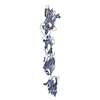
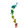
C: von Willebrand factor type A domain protein A: von Willebrand factor type A domain protein D: von Willebrand factor type A domain protein B: von Willebrand factor type A domain protein hetero molecules
Method to determine structure: SAD / Resolution: 2.343→69.139 Å / Cor.coef. Fo:Fc: 0.942 / Cor.coef. Fo:Fc free: 0.909 / SU B: 18.323 / SU ML: 0.196 / Cross valid method: FREE R-VALUE / ESU R: 0.639 / ESU R Free: 0.302 / Details: Hydrogens have not been used
Rfactor
Num. reflection
% reflection
Rfree
0.2396
1787
4.684 %
Rwork
0.1942
36366
-
all
0.196
-
-
obs
-
38153
66.425 %
Solvent computation
Ion probe radii: 0.8 Å / Shrinkage radii: 0.8 Å / VDW probe radii: 1.2 Å / Solvent model: MASK BULK SOLVENT
Displacement parameters
Biso mean: 62.857 Å2
Baniso -1
Baniso -2
Baniso -3
1-
0.747 Å2
-0.162 Å2
-1.082 Å2
2-
-
-0.291 Å2
0.637 Å2
3-
-
-
-0.327 Å2
Refinement step
Cycle: LAST / Resolution: 2.343→69.139 Å
Protein
Nucleic acid
Ligand
Solvent
Total
Num. atoms
7581
0
63
114
7758
Refine LS restraints
Refine-ID
Type
Dev ideal
Dev ideal target
Number
X-RAY DIFFRACTION
r_bond_refined_d
0.011
0.012
7784
X-RAY DIFFRACTION
r_angle_refined_deg
1.82
1.665
10533
X-RAY DIFFRACTION
r_dihedral_angle_1_deg
7.647
5
963
X-RAY DIFFRACTION
r_dihedral_angle_2_deg
40.611
24.684
380
X-RAY DIFFRACTION
r_dihedral_angle_3_deg
17.108
15
1402
X-RAY DIFFRACTION
r_dihedral_angle_4_deg
16.95
15
30
X-RAY DIFFRACTION
r_chiral_restr
0.121
0.2
1059
X-RAY DIFFRACTION
r_gen_planes_refined
0.009
0.02
5760
X-RAY DIFFRACTION
r_nbd_refined
0.211
0.2
2583
X-RAY DIFFRACTION
r_nbtor_refined
0.308
0.2
5227
X-RAY DIFFRACTION
r_xyhbond_nbd_refined
0.143
0.2
252
X-RAY DIFFRACTION
r_symmetry_nbd_refined
0.209
0.2
27
X-RAY DIFFRACTION
r_symmetry_xyhbond_nbd_refined
0.128
0.2
6
X-RAY DIFFRACTION
r_mcbond_it
3.226
3.269
3873
X-RAY DIFFRACTION
r_mcangle_it
5.356
4.883
4829
X-RAY DIFFRACTION
r_scbond_it
4.206
3.622
3911
X-RAY DIFFRACTION
r_scangle_it
6.603
5.267
5704
X-RAY DIFFRACTION
r_lrange_it
10.855
61.109
30786
X-RAY DIFFRACTION
r_ncsr_local_group_1
0.13
0.05
14045
Refine LS restraints NCS
Ens-ID
Dom-ID
Auth asym-ID
Refine-ID
Type
Rms dev position (Å)
Weight position
1
1
A
X-RAY DIFFRACTION
Localncs
0.13009
0.05007
1
2
B
X-RAY DIFFRACTION
Localncs
0.13009
0.05007
LS refinement shell
Resolution (Å)
Rfactor Rfree
Num. reflection Rfree
Rfactor Rwork
Num. reflection Rwork
Refine-ID
% reflection obs (%)
2.343-2.404
0.384
7
0.255
203
X-RAY DIFFRACTION
4.9775
2.404-2.47
0.349
28
0.303
338
X-RAY DIFFRACTION
8.8278
2.47-2.541
0.337
29
0.308
535
X-RAY DIFFRACTION
14.1071
2.541-2.62
0.385
50
0.311
888
X-RAY DIFFRACTION
23.9653
2.62-2.705
0.359
63
0.282
1328
X-RAY DIFFRACTION
36.586
2.705-2.8
0.314
98
0.282
2002
X-RAY DIFFRACTION
56.8182
2.8-2.906
0.355
125
0.286
2903
X-RAY DIFFRACTION
86.7125
2.906-3.024
0.315
148
0.26
3168
X-RAY DIFFRACTION
96.9591
3.024-3.159
0.274
145
0.242
3040
X-RAY DIFFRACTION
97.1333
3.159-3.312
0.298
173
0.227
2859
X-RAY DIFFRACTION
97.5233
3.312-3.491
0.278
138
0.215
2758
X-RAY DIFFRACTION
97.7388
3.491-3.703
0.245
118
0.215
2636
X-RAY DIFFRACTION
97.8678
3.703-3.958
0.256
102
0.198
2471
X-RAY DIFFRACTION
98.0564
3.958-4.274
0.222
114
0.167
2305
X-RAY DIFFRACTION
98.2934
4.274-4.681
0.162
110
0.134
2137
X-RAY DIFFRACTION
98.5526
4.681-5.231
0.17
96
0.12
1906
X-RAY DIFFRACTION
98.7179
5.231-6.036
0.199
86
0.153
1704
X-RAY DIFFRACTION
99.1141
6.036-7.382
0.216
60
0.162
1445
X-RAY DIFFRACTION
99.0132
7.382-10.398
0.15
66
0.138
1123
X-RAY DIFFRACTION
99.4979
10.398-69.139
0.195
31
0.175
617
X-RAY DIFFRACTION
99.2343
Refinement TLS params.
Method: refined / Refine-ID: X-RAY DIFFRACTION
ID
L11 (°2)
L12 (°2)
L13 (°2)
L22 (°2)
L23 (°2)
L33 (°2)
S11 (Å °)
S12 (Å °)
S13 (Å °)
S21 (Å °)
S22 (Å °)
S23 (Å °)
S31 (Å °)
S32 (Å °)
S33 (Å °)
T11 (Å2)
T12 (Å2)
T13 (Å2)
T22 (Å2)
T23 (Å2)
T33 (Å2)
Origin x (Å)
Origin y (Å)
Origin z (Å)
1
0.6996
0.5629
-0.0864
0.8464
0.3722
0.5129
-0.1239
-0.2682
-0.2927
-0.2556
-0.1205
-0.0826
-0.1421
0.1164
0.2443
0.2815
0.0865
0.1152
0.3031
0.0959
0.3951
-18.522
-20.755
-45.247
2
0.0138
0.0352
0.0741
1.3393
1.5805
2.5579
-0.006
-0.0255
-0.0449
-0.0397
-0.0628
0.0233
-0.1418
-0.0298
0.0688
0.1771
-0.0633
0.0023
0.5627
0.0515
0.2071
-21.7014
-0.7722
-25.3065
3
0.6181
0.1164
-0.2694
1.085
1.0555
1.3926
0.0753
0.1989
0.0412
0.1233
0.0654
-0.16
0.0886
-0.2856
-0.1406
0.2281
-0.0155
0.04
0.5119
0.026
0.2186
-19.0863
12.8851
9.2544
4
0.0154
0.1242
-0.018
1.4834
0.1974
1.634
0.0293
0.0485
-0.0428
0.3984
0.118
-0.4267
-0.5267
-0.718
-0.1473
0.4389
0.2004
-0.0189
0.5601
0.005
0.1702
-25.0121
31.4398
41.0992
5
0.1869
-0.0028
-0.246
0.6871
0.3029
0.4598
0.032
0.0305
0.0244
0.079
0.0026
-0.0571
0.0262
-0.0225
-0.0346
0.3428
0.0148
0.0064
0.2865
-0.029
0.3354
2.055
12.153
-1.187
6
0.1509
0.0858
0.0091
1.0922
1.4597
2.0855
0.0247
0.0143
0.0557
0.0435
-0.0309
-0.0466
0.1223
-0.0776
0.0061
0.3708
-0.0177
0.0403
0.2876
-0.0327
0.2929
9.6419
-11.0382
-19.5048
7
0.317
0.1177
-0.0724
0.7825
0.872
1.1377
0.0657
-0.032
0.0435
0.0095
-0.0194
-0.0137
0.0476
-0.0436
-0.0463
0.3725
0.0367
0.0291
0.2779
-0.0422
0.3079
9.7786
-24.956
-54.0289
8
0.0174
0.1136
-0.0408
1.8775
-0.4571
1.267
-0.0144
-0.0284
0.0457
-0.2341
0.0811
-0.0549
0.2176
-0.085
-0.0668
0.3987
-0.0089
0.0171
0.2836
-0.0432
0.3057
1.9871
-44.686
-85.7095
Refinement TLS group
ID
Refine-ID
Refine TLS-ID
Selection
Auth asym-ID
Auth seq-ID
1
X-RAY DIFFRACTION
1
ALL
C
40 - 69
2
X-RAY DIFFRACTION
1
ALL
B
358 - 499
3
X-RAY DIFFRACTION
2
ALL
B
500 - 604
4
X-RAY DIFFRACTION
3
ALL
B
605 - 710
5
X-RAY DIFFRACTION
4
ALL
B
711 - 821
6
X-RAY DIFFRACTION
5
ALL
D
40 - 69
7
X-RAY DIFFRACTION
5
ALL
A
358 - 499
8
X-RAY DIFFRACTION
6
ALL
A
500 - 604
9
X-RAY DIFFRACTION
7
ALL
A
605 - 710
10
X-RAY DIFFRACTION
8
ALL
A
711 - 823
+
About Yorodumi
-
News
-
Feb 9, 2022. New format data for meta-information of EMDB entries
New format data for meta-information of EMDB entries
Version 3 of the EMDB header file is now the official format.
The previous official version 1.9 will be removed from the archive.
In the structure databanks used in Yorodumi, some data are registered as the other names, "COVID-19 virus" and "2019-nCoV". Here are the details of the virus and the list of structure data.
Jan 31, 2019. EMDB accession codes are about to change! (news from PDBe EMDB page)
EMDB accession codes are about to change! (news from PDBe EMDB page)
The allocation of 4 digits for EMDB accession codes will soon come to an end. Whilst these codes will remain in use, new EMDB accession codes will include an additional digit and will expand incrementally as the available range of codes is exhausted. The current 4-digit format prefixed with “EMD-” (i.e. EMD-XXXX) will advance to a 5-digit format (i.e. EMD-XXXXX), and so on. It is currently estimated that the 4-digit codes will be depleted around Spring 2019, at which point the 5-digit format will come into force.
The EM Navigator/Yorodumi systems omit the EMD- prefix.
Related info.:Q: What is EMD? / ID/Accession-code notation in Yorodumi/EM Navigator
Yorodumi is a browser for structure data from EMDB, PDB, SASBDB, etc.
This page is also the successor to EM Navigator detail page, and also detail information page/front-end page for Omokage search.
The word "yorodu" (or yorozu) is an old Japanese word meaning "ten thousand". "mi" (miru) is to see.
Related info.:EMDB / PDB / SASBDB / Comparison of 3 databanks / Yorodumi Search / Aug 31, 2016. New EM Navigator & Yorodumi / Yorodumi Papers / Jmol/JSmol / Function and homology information / Changes in new EM Navigator and Yorodumi
 Movie
Movie Controller

 Yorodumi
 Open data
 Basic information
 Components
 Keywords
 Function and homology information
 Streptococcus oralis ATCC 35037 (bacteria)
 X-RAY DIFFRACTION /
  Authors
 India, 1items
  Citation
 Structure visualization
 Downloads & links
 Other downloads
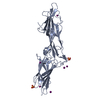

 PDBj
PDBj


 Assembly


 Mass: 556.353 Da / Num. of mol.: 2 / Source method: obtained synthetically / Formula: C20H23N5O4Tb
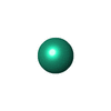
 Mass: 158.925 Da / Num. of mol.: 3 / Source method: obtained synthetically / Formula: Tb
 Mass: 18.015 Da / Num. of mol.: 114 / Source method: isolated from a natural source / Formula: H2O
 Sample preparation
 / Beamline: ID29 / Wavelength: 1.42379 Å
 Processing