 Movie
Movie Controller

[English] 日本語
 Yorodumi
- PDB-7v6f: Structure of Candida albicans Fructose-1,6-bisphosphate aldolase ... -
+ Open data

- Basic information
| Entry | Database: PDB / ID: 7v6f | ||||||
|---|---|---|---|---|---|---|---|
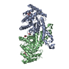
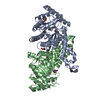
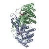
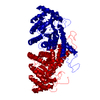
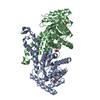
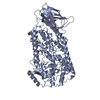
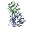
| Title | Structure of Candida albicans Fructose-1,6-bisphosphate aldolase complexed with G3P | ||||||
 Components Components | Fructose-bisphosphate aldolase | ||||||
 Keywords Keywords | LYASE / FBA | ||||||
| Function / homology |  Function and homology information Function and homology informationsymbiont-mediated perturbation of host immune response / hyphal cell wall / melatonin binding / biological process involved in interaction with host / fungal-type cell wall / fructose-bisphosphate aldolase / fructose-bisphosphate aldolase activity / canonical glycolysis / glycolytic process / gluconeogenesis ...symbiont-mediated perturbation of host immune response / hyphal cell wall / melatonin binding / biological process involved in interaction with host / fungal-type cell wall / fructose-bisphosphate aldolase / fructose-bisphosphate aldolase activity / canonical glycolysis / glycolytic process / gluconeogenesis / cellular response to xenobiotic stimulus / cell surface / mitochondrion / zinc ion binding / plasma membrane / cytosol Similarity search - Function | ||||||
| Biological species |  Candida albicans SC5314 (yeast) Candida albicans SC5314 (yeast) | ||||||
| Method |  X-RAY DIFFRACTION / SYNCHROTRON / MOLECULAR REPLACEMENT / Resolution: 2.98 Å X-RAY DIFFRACTION / SYNCHROTRON / MOLECULAR REPLACEMENT / Resolution: 2.98 Å | ||||||
 Authors Authors | Hongxuan, C. / Huang, Y. / Han, C. / Chen, W. / Ren, Y. / Wan, J. | ||||||
| Funding support |  China, 1items China, 1items
| ||||||
 Citation Citation | Journal: J.Med.Chem. / Year: 2022 Title: Structure-Guided Discovery of the Novel Covalent Allosteric Site and Covalent Inhibitors of Fructose-1,6-Bisphosphate Aldolase to Overcome the Azole Resistance of Candidiasis. Authors: Wen, W. / Cao, H. / Huang, Y. / Tu, J. / Wan, C. / Wan, J. / Han, X. / Chen, H. / Liu, J. / Rao, L. / Su, C. / Peng, C. / Sheng, C. / Ren, Y. | ||||||
| History |
|
- Structure visualization
| Structure viewer | Molecule: MolmilJmol/JSmol |
|---|
- Downloads & links
-Download
| PDBx/mmCIF format | 7v6f.cif.gz | 142.4 KB | Display | PDBx/mmCIF format |
|---|---|---|---|---|
| PDB format | pdb7v6f.ent.gz | 109.3 KB | Display | PDB format |
| PDBx/mmJSON format | 7v6f.json.gz | Tree view | PDBx/mmJSON format | |
| Others |  Other downloads Other downloads |
-Validation report
| Arichive directory | https://data.pdbj.org/pub/pdb/validation_reports/v6/7v6fftp://data.pdbj.org/pub/pdb/validation_reports/v6/7v6f | HTTPS FTP |
|---|
-Related structure data
| Related structure data | 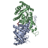 6lnkSC 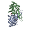 7v6gC S: Starting model for refinement C: citing same article ( |
|---|---|
| Similar structure data |


-Links
 PDBj
PDBj
- Assembly
| Deposited unit | 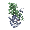
| ||||||||
|---|---|---|---|---|---|---|---|---|---|
| 1 |
| ||||||||
| Unit cell |
|
-Components
| #1: Protein | Mass: 40092.145 Da / Num. of mol.: 2 Source method: isolated from a genetically manipulated source Source: (gene. exp.) Candida albicans SC5314 (yeast) / Strain: SC5314 / Gene: FBA1, CAALFM_C401750CA, CaO19.12088, CaO19.4618 / Production host:  #2: Chemical |   Mass: 65.409 Da / Num. of mol.: 2 / Source method: obtained synthetically / Formula: Zn Mass: 65.409 Da / Num. of mol.: 2 / Source method: obtained synthetically / Formula: Zn#3: Chemical | ChemComp-G3H / |   Mass: 170.058 Da / Num. of mol.: 1 / Source method: obtained synthetically / Formula: C3H7O6P / Feature type: SUBJECT OF INVESTIGATION Mass: 170.058 Da / Num. of mol.: 1 / Source method: obtained synthetically / Formula: C3H7O6P / Feature type: SUBJECT OF INVESTIGATION#4: Water | ChemComp-HOH / |  Mass: 18.015 Da / Num. of mol.: 15 / Source method: isolated from a natural source / Formula: H2O Mass: 18.015 Da / Num. of mol.: 15 / Source method: isolated from a natural source / Formula: H2OHas ligand of interest | Y | |
|---|
-Experimental details
-Experiment
| Experiment | Method: X-RAY DIFFRACTION / Number of used crystals: 1 |
|---|
- Sample preparation
| Crystal | Density Matthews: 2.55 Å3/Da / Density % sol: 51.71 % |
|---|---|
| Crystal grow | Temperature: 291 K / Method: vapor diffusion, hanging drop / Details: 100 mM Hepes pH =7.5, 11% (w/v) PEG 3350 |
-Data collection
| Diffraction | Mean temperature: 100 K / Serial crystal experiment: N |
|---|---|
| Diffraction source | Source: SYNCHROTRON / Site: SSRF / Beamline: BL19U1 / Wavelength: 0.9793 Å |
| Detector | Type: DECTRIS PILATUS 6M / Detector: PIXEL / Date: Jan 2, 2020 |
| Radiation | Protocol: SINGLE WAVELENGTH / Monochromatic (M) / Laue (L): M / Scattering type: x-ray |
| Radiation wavelength | Wavelength: 0.9793 Å / Relative weight: 1 |
| Reflection | Resolution: 2.885→43.68 Å / Num. obs: 18393 / % possible obs: 99.49 % / Redundancy: 6.7 % / CC1/2: 0.999 / Net I/σ(I): 22.7 |
| Reflection shell | Resolution: 2.885→2.988 Å / CC1/2: 0.999 |
- Processing
| Software |
| |||||||||||||||||||||||||||||||||||||||||||||
|---|---|---|---|---|---|---|---|---|---|---|---|---|---|---|---|---|---|---|---|---|---|---|---|---|---|---|---|---|---|---|---|---|---|---|---|---|---|---|---|---|---|---|---|---|---|---|
| Refinement | Method to determine structure: MOLECULAR REPLACEMENT Starting model: 6LNK Resolution: 2.98→43.68 Å / Cor.coef. Fo:Fc: 0.942 / Cor.coef. Fo:Fc free: 0.91 / SU B: 27.549 / SU ML: 0.472 / Cross valid method: FREE R-VALUE / σ(F): 0 / ESU R Free: 0.487 / Stereochemistry target values: MAXIMUM LIKELIHOOD / Details: U VALUES : REFINED INDIVIDUALLY
| |||||||||||||||||||||||||||||||||||||||||||||
| Solvent computation | Ion probe radii: 0.8 Å / Shrinkage radii: 0.8 Å / VDW probe radii: 1.2 Å / Solvent model: MASK | |||||||||||||||||||||||||||||||||||||||||||||
| Displacement parameters | Biso max: 164.63 Å2 / Biso mean: 55.607 Å2 / Biso min: 12.56 Å2
| |||||||||||||||||||||||||||||||||||||||||||||
| Refinement step | Cycle: final / Resolution: 2.98→43.68 Å
| |||||||||||||||||||||||||||||||||||||||||||||
| Refine LS restraints |
| |||||||||||||||||||||||||||||||||||||||||||||
| LS refinement shell | Resolution: 2.984→3.062 Å / Rfactor Rfree error: 0 / Total num. of bins used: 20
|
