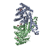
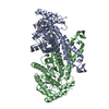
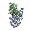
Entry Database : PDB / ID : 5gk7Title Structure of E.Coli fructose 1,6-bisphosphate aldolase bound to Tris Fructose-bisphosphate aldolase class 2 Keywords / / / Function / homology Function Domain/homology Component
/ / / / / / / / / / / / / / / / Biological species Escherichia coli (E. coli)Method / / / Resolution : 1.8 Å Authors Tran, T.H. / Huynh, K.H. / Ho, T.H. / Kang, L.W. Journal : To Be Published Title : Structure of E.Coli fructose 1,6-bisphosphate aldolase, Tris bound formAuthors : Tran, T.H. / Huynh, K.H. / Ho, T.H. / Kang, L.W. History Deposition Jul 3, 2016 Deposition site / Processing site Revision 1.0 Jul 5, 2017 Provider / Type Revision 1.1 Nov 8, 2023 Group Data collection / Database references ... Data collection / Database references / Derived calculations / Refinement description Category chem_comp_atom / chem_comp_bond ... chem_comp_atom / chem_comp_bond / database_2 / pdbx_initial_refinement_model / refine_hist / struct_conn / struct_conn_type Item _database_2.pdbx_DOI / _database_2.pdbx_database_accession ... _database_2.pdbx_DOI / _database_2.pdbx_database_accession / _refine_hist.d_res_low / _struct_conn.conn_type_id / _struct_conn.id / _struct_conn.pdbx_dist_value / _struct_conn.pdbx_leaving_atom_flag / _struct_conn.ptnr1_auth_asym_id / _struct_conn.ptnr1_auth_comp_id / _struct_conn.ptnr1_auth_seq_id / _struct_conn.ptnr1_label_asym_id / _struct_conn.ptnr1_label_atom_id / _struct_conn.ptnr1_label_comp_id / _struct_conn.ptnr1_label_seq_id / _struct_conn.ptnr2_auth_asym_id / _struct_conn.ptnr2_auth_comp_id / _struct_conn.ptnr2_auth_seq_id / _struct_conn.ptnr2_label_asym_id / _struct_conn.ptnr2_label_atom_id / _struct_conn.ptnr2_label_comp_id / _struct_conn.ptnr2_label_seq_id / _struct_conn_type.id Revision 1.2 Oct 23, 2024 Group / Category / pdbx_modification_feature
Show all Show less
 Movie
Movie Controller

 Yorodumi
 Open data
 Basic information
 Components
 Keywords
 Function and homology information
 X-RAY DIFFRACTION /
  Authors
 Citation
 Structure visualization
 Downloads & links
 Other downloads
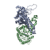


 PDBj
PDBj
 Assembly


 Mass: 92.094 Da / Num. of mol.: 1 / Source method: obtained synthetically / Formula: C3H8O3
 Mass: 122.143 Da / Num. of mol.: 2 / Source method: obtained synthetically / Formula: C4H12NO3 / Comment: pH buffer*YM
 Mass: 106.120 Da / Num. of mol.: 1 / Source method: obtained synthetically / Formula: C4H10O3
 Mass: 65.409 Da / Num. of mol.: 2 / Source method: obtained synthetically / Formula: Zn
 Sample preparation
 / Beamline: 5C (4A) / Wavelength: 0.979 Å
 Processing