 Movie
Movie Controller

[English] 日本語
 Yorodumi
- PDB-5gk8: Structure of E.Coli fructose 1,6-bisphosphate aldolase, Acetate b... -
+ Open data

- Basic information
| Entry | Database: PDB / ID: 5gk8 | ||||||
|---|---|---|---|---|---|---|---|
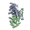
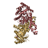
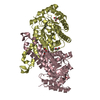
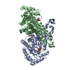
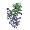
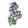
| Title | Structure of E.Coli fructose 1,6-bisphosphate aldolase, Acetate bound form | ||||||
 Components Components | Fructose-bisphosphate aldolase class 2 | ||||||
 Keywords Keywords | LYASE / FBA Acetate bound | ||||||
| Function / homology |  Function and homology information Function and homology informationfructose-bisphosphate aldolase / fructose-bisphosphate aldolase activity / glycolytic process / gluconeogenesis / protein homodimerization activity / zinc ion binding / identical protein binding / cytosol Similarity search - Function | ||||||
| Biological species |  | ||||||
| Method |  X-RAY DIFFRACTION / SYNCHROTRON / MOLECULAR REPLACEMENT / Resolution: 2.002 Å X-RAY DIFFRACTION / SYNCHROTRON / MOLECULAR REPLACEMENT / Resolution: 2.002 Å | ||||||
 Authors Authors | Tran, T.H. / Huynh, K.H. / Ho, T.H. / Kang, L.W. | ||||||
 Citation Citation | Journal: To Be Published Title: Structure of E.Coli fructose 1,6-bisphosphate aldolase, Acetate bound form Authors: Tran, T.H. / Huynh, K.H. / Ho, T.H. / Kang, L.W. | ||||||
| History |
|
- Structure visualization
| Structure viewer | Molecule: MolmilJmol/JSmol |
|---|
- Downloads & links
-Download
| PDBx/mmCIF format | 5gk8.cif.gz | 149.5 KB | Display | PDBx/mmCIF format |
|---|---|---|---|---|
| PDB format | pdb5gk8.ent.gz | 115 KB | Display | PDB format |
| PDBx/mmJSON format | 5gk8.json.gz | Tree view | PDBx/mmJSON format | |
| Others |  Other downloads Other downloads |
-Validation report
| Arichive directory | https://data.pdbj.org/pub/pdb/validation_reports/gk/5gk8ftp://data.pdbj.org/pub/pdb/validation_reports/gk/5gk8 | HTTPS FTP |
|---|
-Related structure data
| Related structure data | 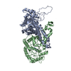 1dosS S: Starting model for refinement |
|---|---|
| Similar structure data |

-Links
 PDBj
PDBj
- Assembly
| Deposited unit | 
| ||||||||
|---|---|---|---|---|---|---|---|---|---|
| 1 |
| ||||||||
| Unit cell |
|
-Components
-Protein , 1 types, 2 molecules AB
| #1: Protein | Mass: 39191.152 Da / Num. of mol.: 2 Source method: isolated from a genetically manipulated source Source: (gene. exp.) Strain: K12 / Gene: fbaA, fba, fda, b2925, JW2892 / Production host: |
|---|
-Non-polymers , 5 types, 365 molecules 


| #2: Chemical | ChemComp-GOL /  Mass: 92.094 Da / Num. of mol.: 4 / Source method: obtained synthetically / Formula: C3H8O3 Mass: 92.094 Da / Num. of mol.: 4 / Source method: obtained synthetically / Formula: C3H8O3#3: Chemical |  Mass: 59.044 Da / Num. of mol.: 2 / Source method: obtained synthetically / Formula: C2H3O2 Mass: 59.044 Da / Num. of mol.: 2 / Source method: obtained synthetically / Formula: C2H3O2#4: Chemical | ChemComp-ZN /  Mass: 65.409 Da / Num. of mol.: 4 / Source method: obtained synthetically / Formula: Zn Mass: 65.409 Da / Num. of mol.: 4 / Source method: obtained synthetically / Formula: Zn#5: Chemical | ChemComp-PEG / |  Mass: 106.120 Da / Num. of mol.: 1 / Source method: obtained synthetically / Formula: C4H10O3 Mass: 106.120 Da / Num. of mol.: 1 / Source method: obtained synthetically / Formula: C4H10O3#6: Water | ChemComp-HOH / | Mass: 18.015 Da / Num. of mol.: 354 / Source method: isolated from a natural source / Formula: H2O |
|---|
-Experimental details
-Experiment
| Experiment | Method: X-RAY DIFFRACTION / Number of used crystals: 1 |
|---|
- Sample preparation
| Crystal | Density Matthews: 2.31 Å3/Da / Density % sol: 46.77 % |
|---|---|
| Crystal grow | Temperature: 287 K / Method: vapor diffusion, hanging drop / pH: 7 Details: 0.2 M ammonium acetate, 5% fructose 1,6-bisphosphatase, 0.1 M Tris pH 7.0, and 15% PEG 4000 PH range: 5.6-7.0 |
-Data collection
| Diffraction | Mean temperature: 100 K |
|---|---|
| Diffraction source | Source: SYNCHROTRON / Site: PAL/PLS  / Beamline: 5C (4A) / Wavelength: 0.979 Å / Beamline: 5C (4A) / Wavelength: 0.979 Å |
| Detector | Type: ADSC QUANTUM 315r / Detector: CCD / Date: Jul 10, 2014 |
| Radiation | Monochromator: DCM Si (111) Crystal / Protocol: MAD / Monochromatic (M) / Laue (L): M / Scattering type: x-ray |
| Radiation wavelength | Wavelength: 0.979 Å / Relative weight: 1 |
| Reflection | Resolution: 2→50 Å / Num. obs: 49638 / % possible obs: 100 % / Redundancy: 6.3 % / Rmerge(I) obs: 0.091 / Net I/σ(I): 40.2 |
| Reflection shell | Resolution: 2→2.05 Å / Redundancy: 6.3 % / Rmerge(I) obs: 0.359 / Mean I/σ(I) obs: 7.6 / % possible all: 100 |
- Processing
| Software |
| ||||||||||||||||||||||||
|---|---|---|---|---|---|---|---|---|---|---|---|---|---|---|---|---|---|---|---|---|---|---|---|---|---|
| Refinement | Method to determine structure: MOLECULAR REPLACEMENT Starting model: 1DOS Resolution: 2.002→36.605 Å / SU ML: 0.18 / Cross valid method: NONE / σ(F): 1.34 / Phase error: 19.21
| ||||||||||||||||||||||||
| Solvent computation | Shrinkage radii: 0.9 Å / VDW probe radii: 1.11 Å | ||||||||||||||||||||||||
| Displacement parameters | Biso max: 90.78 Å2 / Biso mean: 25.0623 Å2 / Biso min: 9.98 Å2 | ||||||||||||||||||||||||
| Refinement step | Cycle: final / Resolution: 2.002→36.605 Å
|
