 Movie
Movie Controller

[English] 日本語
 Yorodumi
- PDB-7v5z: Crystal structure of heterotetrameric complex of Sa2YoeB-Sa2YefM ... -
+ Open data

- Basic information
| Entry | Database: PDB / ID: 7v5z | ||||||
|---|---|---|---|---|---|---|---|
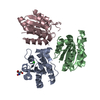
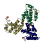
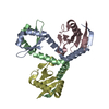
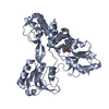
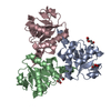
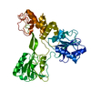
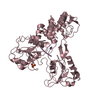
| Title | Crystal structure of heterotetrameric complex of Sa2YoeB-Sa2YefM toxin-antitoxin from Staphylococcus aureus | ||||||
 Components Components |
| ||||||
 Keywords Keywords | ANTITOXIN / TOXIN/ANTITOXIN | ||||||
| Function / homology |  Function and homology information Function and homology informationRNA catabolic process / endonuclease activity / sequence-specific DNA binding / DNA-binding transcription factor activity / negative regulation of DNA-templated transcription / hydrolase activity / regulation of DNA-templated transcription Similarity search - Function | ||||||
| Biological species |   Staphylococcus aureus (bacteria) Staphylococcus aureus (bacteria) | ||||||
| Method |  X-RAY DIFFRACTION / SYNCHROTRON / MOLECULAR REPLACEMENT / Resolution: 1.99 Å X-RAY DIFFRACTION / SYNCHROTRON / MOLECULAR REPLACEMENT / Resolution: 1.99 Å | ||||||
 Authors Authors | Xue, L. / Khan, M.H. / Yue, J. | ||||||
| Funding support |  China, 1items China, 1items
| ||||||
 Citation Citation | Journal: J.Biol.Chem. / Year: 2022 Title: The two paralogous copies of the YoeB-YefM toxin-antitoxin module in Staphylococcus aureus differ in DNA binding and recognition patterns. Authors: Xue, L. / Khan, M.H. / Yue, J. / Zhu, Z. / Niu, L. | ||||||
| History |
|
- Structure visualization
| Structure viewer | Molecule: MolmilJmol/JSmol |
|---|
- Downloads & links
-Download
| PDBx/mmCIF format | 7v5z.cif.gz | 107.4 KB | Display | PDBx/mmCIF format |
|---|---|---|---|---|
| PDB format | pdb7v5z.ent.gz | 66.4 KB | Display | PDB format |
| PDBx/mmJSON format | 7v5z.json.gz | Tree view | PDBx/mmJSON format | |
| Others |  Other downloads Other downloads |
-Validation report
| Arichive directory | https://data.pdbj.org/pub/pdb/validation_reports/v5/7v5zftp://data.pdbj.org/pub/pdb/validation_reports/v5/7v5z | HTTPS FTP |
|---|
-Related structure data
| Related structure data |  7v5ySC  7v6wC S: Starting model for refinement C: citing same article ( |
|---|---|
| Similar structure data |

-Links
 PDBj
PDBj- Assembly
| Deposited unit | 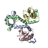
| ||||||||||||
|---|---|---|---|---|---|---|---|---|---|---|---|---|---|
| 1 |
| ||||||||||||
| Unit cell |
|
-Components
| #1: Protein | Mass: 9783.692 Da / Num. of mol.: 2 Source method: isolated from a genetically manipulated source Source: (gene. exp.) Staphylococcus aureus (strain NCTC 8325 / PS 47) (bacteria)Strain: NCTC 8325 / PS 47 / Gene: SAOUHSC_02757 / Production host: #2: Protein | Mass: 10670.266 Da / Num. of mol.: 2 Source method: isolated from a genetically manipulated source Source: (gene. exp.) Staphylococcus aureus (strain NCTC 8325 / PS 47) (bacteria)Strain: NCTC 8325 / PS 47 / Gene: SAOUHSC_02756 / Production host: #3: Water | ChemComp-HOH / |  Mass: 18.015 Da / Num. of mol.: 240 / Source method: isolated from a natural source / Formula: H2O Mass: 18.015 Da / Num. of mol.: 240 / Source method: isolated from a natural source / Formula: H2O |
|---|
-Experimental details
-Experiment
| Experiment | Method: X-RAY DIFFRACTION / Number of used crystals: 1 |
|---|
- Sample preparation
| Crystal | Density Matthews: 2.01 Å3/Da / Density % sol: 38.67 % |
|---|---|
| Crystal grow | Temperature: 289.15 K / Method: vapor diffusion, sitting drop / pH: 7 Details: 0.2 M Succinic acid pH 7.0, 20% w/v Polyethylene glycol 3350 |
-Data collection
| Diffraction | Mean temperature: 100 K / Serial crystal experiment: N |
|---|---|
| Diffraction source | Source: SYNCHROTRON / Site: SSRF / Beamline: BL19U1 / Wavelength: 0.97853 Å |
| Detector | Type: DECTRIS PILATUS3 6M / Detector: PIXEL / Date: Nov 9, 2019 |
| Radiation | Protocol: SINGLE WAVELENGTH / Monochromatic (M) / Laue (L): M / Scattering type: x-ray |
| Radiation wavelength | Wavelength: 0.97853 Å / Relative weight: 1 |
| Reflection | Resolution: 1.99→50 Å / Num. obs: 23229 / % possible obs: 99.8 % / Redundancy: 12.5 % / Biso Wilson estimate: 29.57 Å2 / CC1/2: 0.991 / Net I/σ(I): 12.1 |
| Reflection shell | Resolution: 1.99→2.03 Å / Redundancy: 10.4 % / Num. unique obs: 1152 / CC1/2: 0.768 / % possible all: 99.7 |
- Processing
| Software |
| |||||||||||||||||||||||||||||||||||||||||||||||||||||||||||||||
|---|---|---|---|---|---|---|---|---|---|---|---|---|---|---|---|---|---|---|---|---|---|---|---|---|---|---|---|---|---|---|---|---|---|---|---|---|---|---|---|---|---|---|---|---|---|---|---|---|---|---|---|---|---|---|---|---|---|---|---|---|---|---|---|---|
| Refinement | Method to determine structure: MOLECULAR REPLACEMENT Starting model: 7V5Y Resolution: 1.99→33.76 Å / SU ML: 0.2008 / Cross valid method: FREE R-VALUE / σ(F): 1.34 / Phase error: 20.8523 Stereochemistry target values: GeoStd + Monomer Library + CDL v1.2
| |||||||||||||||||||||||||||||||||||||||||||||||||||||||||||||||
| Solvent computation | Shrinkage radii: 0.9 Å / VDW probe radii: 1.11 Å / Solvent model: FLAT BULK SOLVENT MODEL | |||||||||||||||||||||||||||||||||||||||||||||||||||||||||||||||
| Displacement parameters | Biso mean: 31.44 Å2 | |||||||||||||||||||||||||||||||||||||||||||||||||||||||||||||||
| Refinement step | Cycle: LAST / Resolution: 1.99→33.76 Å
| |||||||||||||||||||||||||||||||||||||||||||||||||||||||||||||||
| Refine LS restraints |
| |||||||||||||||||||||||||||||||||||||||||||||||||||||||||||||||
| LS refinement shell |
|
