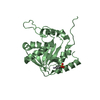
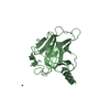
 Movie
Movie Controller


+ Open data

- Basic information
| Entry | Database: PDB / ID: 7v39 | |||||||||
|---|---|---|---|---|---|---|---|---|---|---|
| Title | Crystal structure of NP exonuclease-PCMB complex | |||||||||
 Components Components | Nucleoprotein | |||||||||
 Keywords Keywords | VIRAL PROTEIN / DEDDh exonuclease / NP exonuclease / anti-viral drug / PCMPS / PCMB | |||||||||
| Function / homology |  Function and homology information Function and homology informationsymbiont-mediated suppression of host cytoplasmic pattern recognition receptor signaling pathway via inhibition of IKBKE activity / RNA-templated viral transcription / negative stranded viral RNA replication / helical viral capsid / 3'-5' exonuclease activity / viral nucleocapsid / Hydrolases; Acting on ester bonds; Exoribonucleases producing 5'-phosphomonoesters / symbiont-mediated suppression of host cytoplasmic pattern recognition receptor signaling pathway via inhibition of IRF3 activity / host cell cytoplasm / ribonucleoprotein complex ...symbiont-mediated suppression of host cytoplasmic pattern recognition receptor signaling pathway via inhibition of IKBKE activity / RNA-templated viral transcription / negative stranded viral RNA replication / helical viral capsid / 3'-5' exonuclease activity / viral nucleocapsid / Hydrolases; Acting on ester bonds; Exoribonucleases producing 5'-phosphomonoesters / symbiont-mediated suppression of host cytoplasmic pattern recognition receptor signaling pathway via inhibition of IRF3 activity / host cell cytoplasm / ribonucleoprotein complex / RNA binding / metal ion binding / identical protein binding Similarity search - Function | |||||||||
| Biological species |  Lassa virus Lassa virus | |||||||||
| Method |  X-RAY DIFFRACTION / SYNCHROTRON / MOLECULAR REPLACEMENT / Resolution: 2.2 Å X-RAY DIFFRACTION / SYNCHROTRON / MOLECULAR REPLACEMENT / Resolution: 2.2 Å | |||||||||
 Authors Authors | Hsiao, Y.Y. / Huang, K.W. | |||||||||
| Funding support |  Taiwan, 2items Taiwan, 2items
| |||||||||
 Citation Citation | Journal: Jacs Au / Year: 2021 Title: Targeted Covalent Inhibitors Allosterically Deactivate the DEDDh Lassa Fever Virus NP Exonuclease from Alternative Distal Sites. Authors: Huang, K.W. / Chen, J.W. / Hua, T.Y. / Chu, Y.Y. / Chiu, T.Y. / Liu, J.Y. / Tu, C.I. / Hsu, K.C. / Kao, Y.T. / Chu, J.W. / Hsiao, Y.Y. | |||||||||
| History |
|
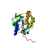
- Structure visualization
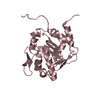
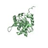
| Structure viewer | Molecule: MolmilJmol/JSmol |
|---|
- Downloads & links
-Download
| PDBx/mmCIF format | 7v39.cif.gz | 189.2 KB | Display | PDBx/mmCIF format |
|---|---|---|---|---|
| PDB format | pdb7v39.ent.gz | 147.2 KB | Display | PDB format |
| PDBx/mmJSON format | 7v39.json.gz | Tree view | PDBx/mmJSON format | |
| Others |  Other downloads Other downloads |
-Validation report
| Arichive directory | https://data.pdbj.org/pub/pdb/validation_reports/v3/7v39ftp://data.pdbj.org/pub/pdb/validation_reports/v3/7v39 | HTTPS FTP |
|---|
-Related structure data
| Related structure data |  7v37SC  7v38C 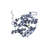 7v3aC 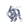 7v3bC 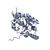 7v3cC S: Starting model for refinement C: citing same article ( |
|---|---|
| Similar structure data |


-Links
 PDBj
PDBj- Assembly
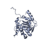
| Deposited unit | 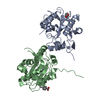
| ||||||||||
|---|---|---|---|---|---|---|---|---|---|---|---|
| 1 | 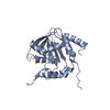
| ||||||||||
| 2 | 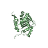
| ||||||||||
| Unit cell |
|
-Components
| #1: Protein | Mass: 27693.641 Da / Num. of mol.: 2 Source method: isolated from a genetically manipulated source Source: (gene. exp.) Lassa virus (strain Mouse/Sierra Leone/Josiah/1976)Strain: Mouse/Sierra Leone/Josiah/1976 / Production host:  References: UniProt: P13699, Hydrolases; Acting on ester bonds; Exoribonucleases producing 5'-phosphomonoesters #2: Chemical |   Mass: 65.409 Da / Num. of mol.: 2 / Source method: obtained synthetically / Formula: Zn / Feature type: SUBJECT OF INVESTIGATION Mass: 65.409 Da / Num. of mol.: 2 / Source method: obtained synthetically / Formula: Zn / Feature type: SUBJECT OF INVESTIGATION#3: Chemical | 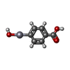  Mass: 338.711 Da / Num. of mol.: 2 / Source method: obtained synthetically / Formula: C7H6HgO3 / Feature type: SUBJECT OF INVESTIGATION Mass: 338.711 Da / Num. of mol.: 2 / Source method: obtained synthetically / Formula: C7H6HgO3 / Feature type: SUBJECT OF INVESTIGATION#4: Water | ChemComp-HOH / |  Mass: 18.015 Da / Num. of mol.: 273 / Source method: isolated from a natural source / Formula: H2O Mass: 18.015 Da / Num. of mol.: 273 / Source method: isolated from a natural source / Formula: H2OHas ligand of interest | Y | |
|---|
-Experimental details
-Experiment
| Experiment | Method: X-RAY DIFFRACTION / Number of used crystals: 1 |
|---|
- Sample preparation
| Crystal | Density Matthews: 2.43 Å3/Da / Density % sol: 49.44 % |
|---|---|
| Crystal grow | Temperature: 293 K / Method: vapor diffusion, hanging drop Details: 0.2 M DL-Malic acid pH 7.0, 20% w/v Polyethylene glycol 3,350, 2mM Sodium p-hydroxymercuribenzoate |
-Data collection
| Diffraction | Mean temperature: 100 K / Serial crystal experiment: N | |||||||||||||||||||||||||||||||||||||||||||||||||||||||||||||||||||||||||||||||||||||||||||||||||||
|---|---|---|---|---|---|---|---|---|---|---|---|---|---|---|---|---|---|---|---|---|---|---|---|---|---|---|---|---|---|---|---|---|---|---|---|---|---|---|---|---|---|---|---|---|---|---|---|---|---|---|---|---|---|---|---|---|---|---|---|---|---|---|---|---|---|---|---|---|---|---|---|---|---|---|---|---|---|---|---|---|---|---|---|---|---|---|---|---|---|---|---|---|---|---|---|---|---|---|---|---|
| Diffraction source | Source: SYNCHROTRON / Site: NSRRC / Beamline: BL15A1 / Wavelength: 1 Å | |||||||||||||||||||||||||||||||||||||||||||||||||||||||||||||||||||||||||||||||||||||||||||||||||||
| Detector | Type: RAYONIX MX300HE / Detector: CCD / Date: Mar 4, 2016 | |||||||||||||||||||||||||||||||||||||||||||||||||||||||||||||||||||||||||||||||||||||||||||||||||||
| Radiation | Protocol: SINGLE WAVELENGTH / Monochromatic (M) / Laue (L): M / Scattering type: x-ray | |||||||||||||||||||||||||||||||||||||||||||||||||||||||||||||||||||||||||||||||||||||||||||||||||||
| Radiation wavelength | Wavelength: 1 Å / Relative weight: 1 | |||||||||||||||||||||||||||||||||||||||||||||||||||||||||||||||||||||||||||||||||||||||||||||||||||
| Reflection | Resolution: 2.2→30 Å / Num. obs: 28303 / % possible obs: 99.9 % / Redundancy: 6.3 % / Biso Wilson estimate: 29.98 Å2 / Rmerge(I) obs: 0.089 / Rpim(I) all: 0.038 / Rrim(I) all: 0.097 / Χ2: 1.941 / Net I/σ(I): 11.8 / Num. measured all: 179251 | |||||||||||||||||||||||||||||||||||||||||||||||||||||||||||||||||||||||||||||||||||||||||||||||||||
| Reflection shell | Diffraction-ID: 1
|
- Processing
| Software |
| ||||||||||||||||||||||||||||||||||||||||||||||||||||||||||||||||||||||||||||||||
|---|---|---|---|---|---|---|---|---|---|---|---|---|---|---|---|---|---|---|---|---|---|---|---|---|---|---|---|---|---|---|---|---|---|---|---|---|---|---|---|---|---|---|---|---|---|---|---|---|---|---|---|---|---|---|---|---|---|---|---|---|---|---|---|---|---|---|---|---|---|---|---|---|---|---|---|---|---|---|---|---|---|
| Refinement | Method to determine structure: MOLECULAR REPLACEMENT Starting model: 7V37 Resolution: 2.2→28.816 Å / SU ML: 0.21 / Cross valid method: THROUGHOUT / σ(F): 1.37 / Phase error: 21.87
| ||||||||||||||||||||||||||||||||||||||||||||||||||||||||||||||||||||||||||||||||
| Solvent computation | Shrinkage radii: 0.9 Å / VDW probe radii: 1.11 Å | ||||||||||||||||||||||||||||||||||||||||||||||||||||||||||||||||||||||||||||||||
| Displacement parameters | Biso max: 236.39 Å2 / Biso mean: 38.577 Å2 / Biso min: 14.99 Å2 | ||||||||||||||||||||||||||||||||||||||||||||||||||||||||||||||||||||||||||||||||
| Refinement step | Cycle: final / Resolution: 2.2→28.816 Å
| ||||||||||||||||||||||||||||||||||||||||||||||||||||||||||||||||||||||||||||||||
| LS refinement shell | Refine-ID: X-RAY DIFFRACTION / Rfactor Rfree error: 0 / % reflection obs: 100 %
| ||||||||||||||||||||||||||||||||||||||||||||||||||||||||||||||||||||||||||||||||
| Refinement TLS params. | Method: refined / Origin x: -36.9261 Å / Origin y: 6.3774 Å / Origin z: -35.6693 Å
| ||||||||||||||||||||||||||||||||||||||||||||||||||||||||||||||||||||||||||||||||
| Refinement TLS group |
|
