 Movie
Movie Controller

[English] 日本語
 Yorodumi
- PDB-7uxu: CryoEM structure of the TIR domain from AbTir in complex with 3AD -
+ Open data

- Basic information
| Entry | Database: PDB / ID: 7uxu | |||||||||
|---|---|---|---|---|---|---|---|---|---|---|
| Title | CryoEM structure of the TIR domain from AbTir in complex with 3AD | |||||||||
 Components Components | Molecular chaperone Tir | |||||||||
 Keywords Keywords | HYDROLASE / 2' cADPR / NADase / Bacterial TIR | |||||||||
| Function / homology |  Function and homology information Function and homology informationNAD+ nucleosidase activity / NAD+ catabolic process / signal transduction Similarity search - Function | |||||||||
| Biological species |  Acinetobacter baumannii (bacteria) Acinetobacter baumannii (bacteria) | |||||||||
| Method | ELECTRON MICROSCOPY / helical reconstruction / cryo EM / Resolution: 2.74 Å | |||||||||
 Authors Authors | Li, S. / Nanson, J.D. / Manik, M.K. / Gu, W. / Landsberg, M.J. / Ve, T. / Kobe, B. | |||||||||
| Funding support |  Australia, 2items Australia, 2items
| |||||||||
 Citation Citation | Journal: Science / Year: 2022 Title: Cyclic ADP ribose isomers: Production, chemical structures, and immune signaling. Authors: Mohammad K Manik / Yun Shi / Sulin Li / Mark A Zaydman / Neha Damaraju / Samuel Eastman / Thomas G Smith / Weixi Gu / Veronika Masic / Tamim Mosaiab / James S Weagley / Steven J Hancock / ...Authors: Mohammad K Manik / Yun Shi / Sulin Li / Mark A Zaydman / Neha Damaraju / Samuel Eastman / Thomas G Smith / Weixi Gu / Veronika Masic / Tamim Mosaiab / James S Weagley / Steven J Hancock / Eduardo Vasquez / Lauren Hartley-Tassell / Nestoras Kargios / Natsumi Maruta / Bryan Y J Lim / Hayden Burdett / Michael J Landsberg / Mark A Schembri / Ivan Prokes / Lijiang Song / Murray Grant / Aaron DiAntonio / Jeffrey D Nanson / Ming Guo / Jeffrey Milbrandt / Thomas Ve / Bostjan Kobe /   Abstract: Cyclic adenosine diphosphate (ADP)-ribose (cADPR) isomers are signaling molecules produced by bacterial and plant Toll/interleukin-1 receptor (TIR) domains via nicotinamide adenine dinucleotide ...Cyclic adenosine diphosphate (ADP)-ribose (cADPR) isomers are signaling molecules produced by bacterial and plant Toll/interleukin-1 receptor (TIR) domains via nicotinamide adenine dinucleotide (oxidized form) (NAD) hydrolysis. We show that v-cADPR (2'cADPR) and v2-cADPR (3'cADPR) isomers are cyclized by O-glycosidic bond formation between the ribose moieties in ADPR. Structures of 2'cADPR-producing TIR domains reveal conformational changes that lead to an active assembly that resembles those of Toll-like receptor adaptor TIR domains. Mutagenesis reveals a conserved tryptophan that is essential for cyclization. We show that 3'cADPR is an activator of ThsA effector proteins from the bacterial antiphage defense system termed Thoeris and a suppressor of plant immunity when produced by the effector HopAM1. Collectively, our results reveal the molecular basis of cADPR isomer production and establish 3'cADPR in bacteria as an antiviral and plant immunity-suppressing signaling molecule. | |||||||||
| History |
|
- Structure visualization
| Structure viewer | Molecule: MolmilJmol/JSmol |
|---|
- Downloads & links
-Download
| PDBx/mmCIF format | 7uxu.cif.gz | 265.6 KB | Display | PDBx/mmCIF format |
|---|---|---|---|---|
| PDB format | pdb7uxu.ent.gz | 176.1 KB | Display | PDB format |
| PDBx/mmJSON format | 7uxu.json.gz | Tree view | PDBx/mmJSON format | |
| Others |  Other downloads Other downloads |
-Validation report
| Arichive directory | https://data.pdbj.org/pub/pdb/validation_reports/ux/7uxuftp://data.pdbj.org/pub/pdb/validation_reports/ux/7uxu | HTTPS FTP |
|---|
-Related structure data
| Related structure data | 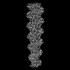 26862MC 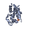 7uwgC 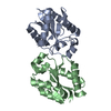 7uxrC 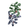 7uxsC  7uxtC M: map data used to model this data C: citing same article ( |
|---|---|
| Similar structure data |
-Links
 PDBj
PDBj
- Assembly
| Deposited unit | 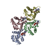
|
|---|---|
| 1 |
|
| Symmetry | Helical symmetry: (Circular symmetry: 1 / Dyad axis: no / N subunits divisor: 1 / Num. of operations: 4 / Rise per n subunits: 17.8 Å / Rotation per n subunits: 174 °) |
-Components
| #1: Protein | Mass: 15614.538 Da / Num. of mol.: 4 Source method: isolated from a genetically manipulated source Source: (gene. exp.) Acinetobacter baumannii (bacteria) / Gene: APD31_10100, H0529_09530 / Production host: #2: Chemical | ChemComp-1O4 / [[( 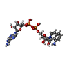  Mass: 686.482 Da / Num. of mol.: 4 / Source method: obtained synthetically / Formula: C24H30N7O13P2 / Feature type: SUBJECT OF INVESTIGATION Mass: 686.482 Da / Num. of mol.: 4 / Source method: obtained synthetically / Formula: C24H30N7O13P2 / Feature type: SUBJECT OF INVESTIGATIONHas ligand of interest | Y | |
|---|
-Experimental details
-Experiment
| Experiment | Method: ELECTRON MICROSCOPY |
|---|---|
| EM experiment | Aggregation state: FILAMENT / 3D reconstruction method: helical reconstruction |
- Sample preparation
| Component | Name: Filament of the AbTIR TIR domain in complex with 3AD / Type: COMPLEX / Entity ID: #1 / Source: RECOMBINANT |
|---|---|
| Source (natural) | Organism: Acinetobacter baumannii (bacteria) |
| Source (recombinant) | Organism: |
| Buffer solution | pH: 7.5 |
| Specimen | Conc.: 2.5 mg/ml / Embedding applied: NO / Shadowing applied: NO / Staining applied: NO / Vitrification applied: YES |
| Specimen support | Grid material: COPPER / Grid type: Quantifoil |
| Vitrification | Instrument: LEICA EM GP / Cryogen name: ETHANE / Humidity: 96 % / Chamber temperature: 281 K |
- Electron microscopy imaging
| Microscopy | Model: JEOL CRYO ARM 300 |
|---|---|
| Electron gun | Electron source:  FIELD EMISSION GUN / Accelerating voltage: 300 kV / Illumination mode: OTHER FIELD EMISSION GUN / Accelerating voltage: 300 kV / Illumination mode: OTHER |
| Electron lens | Mode: OTHER / Nominal defocus max: 4000 nm / Nominal defocus min: 100 nm / Cs: 2.7 mm |
| Image recording | Electron dose: 40.9 e/Å2 / Film or detector model: GATAN K3 (6k x 4k) |
- Processing
| Software |
| ||||||||||||||||||||||||
|---|---|---|---|---|---|---|---|---|---|---|---|---|---|---|---|---|---|---|---|---|---|---|---|---|---|
| EM software |
| ||||||||||||||||||||||||
| CTF correction | Type: PHASE FLIPPING AND AMPLITUDE CORRECTION | ||||||||||||||||||||||||
| Helical symmerty | Angular rotation/subunit: 174.056 ° / Axial rise/subunit: 17.867 Å / Axial symmetry: C1 | ||||||||||||||||||||||||
| 3D reconstruction | Resolution: 2.74 Å / Resolution method: FSC 0.143 CUT-OFF / Num. of particles: 272949 / Symmetry type: HELICAL | ||||||||||||||||||||||||
| Refinement | Cross valid method: NONE Stereochemistry target values: GeoStd + Monomer Library + CDL v1.2 | ||||||||||||||||||||||||
| Displacement parameters | Biso mean: 33.48 Å2 | ||||||||||||||||||||||||
| Refine LS restraints |
|
