 Movie
Movie Controller

[English] 日本語
 Yorodumi
- PDB-7uwg: The crystal structure of the TIR domain-containing protein from A... -
+ Open data

- Basic information
| Entry | Database: PDB / ID: 7uwg | ||||||
|---|---|---|---|---|---|---|---|
| Title | The crystal structure of the TIR domain-containing protein from Acinetobacter baumannii (AbTir) | ||||||
 Components Components | Molecular chaperone Tir | ||||||
 Keywords Keywords | HYDROLASE / 2' cADPR / NADase / Bacterial TIR | ||||||
| Function / homology | NAD+ catabolic process / NAD+ nucleosidase activity / TIR domain / Toll - interleukin 1 - resistance / TIR domain profile. / Toll/interleukin-1 receptor homology (TIR) domain / Toll/interleukin-1 receptor homology (TIR) domain superfamily / signal transduction / Molecular chaperone Tir Function and homology information Function and homology information | ||||||
| Biological species |  Acinetobacter baumannii (bacteria) Acinetobacter baumannii (bacteria) | ||||||
| Method |  X-RAY DIFFRACTION / SYNCHROTRON / MOLECULAR REPLACEMENT / Resolution: 2.16 Å X-RAY DIFFRACTION / SYNCHROTRON / MOLECULAR REPLACEMENT / Resolution: 2.16 Å | ||||||
 Authors Authors | Manik, M.K. / Nanson, J.D. / Ve, T. / Kobe, B. | ||||||
| Funding support |  Australia, 1items Australia, 1items
| ||||||
 Citation Citation | Journal: Science / Year: 2022 Title: Cyclic ADP ribose isomers: Production, chemical structures, and immune signaling. Authors: Mohammad K Manik / Yun Shi / Sulin Li / Mark A Zaydman / Neha Damaraju / Samuel Eastman / Thomas G Smith / Weixi Gu / Veronika Masic / Tamim Mosaiab / James S Weagley / Steven J Hancock / ...Authors: Mohammad K Manik / Yun Shi / Sulin Li / Mark A Zaydman / Neha Damaraju / Samuel Eastman / Thomas G Smith / Weixi Gu / Veronika Masic / Tamim Mosaiab / James S Weagley / Steven J Hancock / Eduardo Vasquez / Lauren Hartley-Tassell / Nestoras Kargios / Natsumi Maruta / Bryan Y J Lim / Hayden Burdett / Michael J Landsberg / Mark A Schembri / Ivan Prokes / Lijiang Song / Murray Grant / Aaron DiAntonio / Jeffrey D Nanson / Ming Guo / Jeffrey Milbrandt / Thomas Ve / Bostjan Kobe /   Abstract: Cyclic adenosine diphosphate (ADP)-ribose (cADPR) isomers are signaling molecules produced by bacterial and plant Toll/interleukin-1 receptor (TIR) domains via nicotinamide adenine dinucleotide ...Cyclic adenosine diphosphate (ADP)-ribose (cADPR) isomers are signaling molecules produced by bacterial and plant Toll/interleukin-1 receptor (TIR) domains via nicotinamide adenine dinucleotide (oxidized form) (NAD) hydrolysis. We show that v-cADPR (2'cADPR) and v2-cADPR (3'cADPR) isomers are cyclized by O-glycosidic bond formation between the ribose moieties in ADPR. Structures of 2'cADPR-producing TIR domains reveal conformational changes that lead to an active assembly that resembles those of Toll-like receptor adaptor TIR domains. Mutagenesis reveals a conserved tryptophan that is essential for cyclization. We show that 3'cADPR is an activator of ThsA effector proteins from the bacterial antiphage defense system termed Thoeris and a suppressor of plant immunity when produced by the effector HopAM1. Collectively, our results reveal the molecular basis of cADPR isomer production and establish 3'cADPR in bacteria as an antiviral and plant immunity-suppressing signaling molecule. | ||||||
| History |
|
- Structure visualization
| Structure viewer | Molecule: MolmilJmol/JSmol |
|---|
- Downloads & links
-Download
| PDBx/mmCIF format | 7uwg.cif.gz | 137.5 KB | Display | PDBx/mmCIF format |
|---|---|---|---|---|
| PDB format | pdb7uwg.ent.gz | 97.1 KB | Display | PDB format |
| PDBx/mmJSON format | 7uwg.json.gz | Tree view | PDBx/mmJSON format | |
| Others |  Other downloads Other downloads |
-Validation report
| Summary document | 7uwg_validation.pdf.gz | 2.1 MB | Display | wwPDB validaton report |
|---|---|---|---|---|
| Full document | 7uwg_full_validation.pdf.gz | 2.1 MB | Display | |
| Data in XML | 7uwg_validation.xml.gz | 22.9 KB | Display | |
| Data in CIF | 7uwg_validation.cif.gz | 32.2 KB | Display | |
| Arichive directory | https://data.pdbj.org/pub/pdb/validation_reports/uw/7uwgftp://data.pdbj.org/pub/pdb/validation_reports/uw/7uwg | HTTPS FTP |
-Related structure data
| Related structure data | 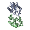 7uxrC 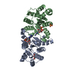 7uxsC  7uxtC 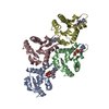 7uxuC 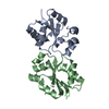 4lqcS S: Starting model for refinement C: citing same article ( |
|---|---|
| Similar structure data |
-Links
 PDBj
PDBj
- Assembly
| Deposited unit | 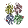
| ||||||||||||
|---|---|---|---|---|---|---|---|---|---|---|---|---|---|
| 1 | 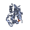
| ||||||||||||
| 2 | 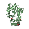
| ||||||||||||
| 3 | 
| ||||||||||||
| 4 | 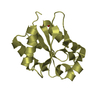
| ||||||||||||
| Unit cell |
|
-Components
| #1: Protein | Mass: 15614.538 Da / Num. of mol.: 4 Source method: isolated from a genetically manipulated source Source: (gene. exp.) Acinetobacter baumannii (bacteria) / Gene: APD31_10100, H0529_09530 / Production host: #2: Chemical | ChemComp-SO4 /   Mass: 96.063 Da / Num. of mol.: 5 / Source method: obtained synthetically / Formula: SO4 / Feature type: SUBJECT OF INVESTIGATION Mass: 96.063 Da / Num. of mol.: 5 / Source method: obtained synthetically / Formula: SO4 / Feature type: SUBJECT OF INVESTIGATION#3: Chemical | ChemComp-P6G / |   Mass: 282.331 Da / Num. of mol.: 1 / Source method: isolated from a natural source / Formula: C12H26O7 / Feature type: SUBJECT OF INVESTIGATION / Comment: precipitant*YM Mass: 282.331 Da / Num. of mol.: 1 / Source method: isolated from a natural source / Formula: C12H26O7 / Feature type: SUBJECT OF INVESTIGATION / Comment: precipitant*YM#4: Water | ChemComp-HOH / |  Mass: 18.015 Da / Num. of mol.: 237 / Source method: isolated from a natural source / Formula: H2O Mass: 18.015 Da / Num. of mol.: 237 / Source method: isolated from a natural source / Formula: H2OHas ligand of interest | Y | |
|---|
-Experimental details
-Experiment
| Experiment | Method: X-RAY DIFFRACTION / Number of used crystals: 1 |
|---|
- Sample preparation
| Crystal | Density Matthews: 2 Å3/Da / Density % sol: 38.54 % |
|---|---|
| Crystal grow | Temperature: 293 K / Method: vapor diffusion, hanging drop / pH: 5.5 Details: 0.1 M Bis-Tris pH 5.5, 0.2 M LiSO4, and 25% PEG 3350 |
-Data collection
| Diffraction | Mean temperature: 100 K / Serial crystal experiment: N |
|---|---|
| Diffraction source | Source: SYNCHROTRON / Site: Australian Synchrotron / Beamline: MX2 / Wavelength: 0.9537 Å |
| Detector | Type: DECTRIS EIGER X 16M / Detector: PIXEL / Date: Feb 14, 2018 |
| Radiation | Protocol: SINGLE WAVELENGTH / Monochromatic (M) / Laue (L): M / Scattering type: x-ray |
| Radiation wavelength | Wavelength: 0.9537 Å / Relative weight: 1 |
| Reflection | Resolution: 2.16→47.86 Å / Num. obs: 25989 / % possible obs: 97.9 % / Redundancy: 3.8 % / Biso Wilson estimate: 25.33 Å2 / CC1/2: 0.99 / Rmerge(I) obs: 0.12 / Rpim(I) all: 0.1 / Rrim(I) all: 0.14 / Net I/σ(I): 7.6 |
| Reflection shell | Resolution: 2.16→2.24 Å / Rmerge(I) obs: 0.66 / Num. unique obs: 2180 / CC1/2: 0.77 / Rpim(I) all: 0.52 |
- Processing
| Software |
| |||||||||||||||||||||||||||||||||||||||||||||||||||||||||||||||||||||||||||||||||||||||||||||||||||||||||
|---|---|---|---|---|---|---|---|---|---|---|---|---|---|---|---|---|---|---|---|---|---|---|---|---|---|---|---|---|---|---|---|---|---|---|---|---|---|---|---|---|---|---|---|---|---|---|---|---|---|---|---|---|---|---|---|---|---|---|---|---|---|---|---|---|---|---|---|---|---|---|---|---|---|---|---|---|---|---|---|---|---|---|---|---|---|---|---|---|---|---|---|---|---|---|---|---|---|---|---|---|---|---|---|---|---|---|
| Refinement | Method to determine structure: MOLECULAR REPLACEMENT Starting model: 4lqc Resolution: 2.16→47.86 Å / SU ML: 0.2665 / Cross valid method: FREE R-VALUE / σ(F): 1.38 / Phase error: 24.1275 Stereochemistry target values: GeoStd + Monomer Library + CDL v1.2
| |||||||||||||||||||||||||||||||||||||||||||||||||||||||||||||||||||||||||||||||||||||||||||||||||||||||||
| Solvent computation | Shrinkage radii: 0.9 Å / VDW probe radii: 1.1 Å / Solvent model: FLAT BULK SOLVENT MODEL | |||||||||||||||||||||||||||||||||||||||||||||||||||||||||||||||||||||||||||||||||||||||||||||||||||||||||
| Displacement parameters | Biso mean: 29.35 Å2 | |||||||||||||||||||||||||||||||||||||||||||||||||||||||||||||||||||||||||||||||||||||||||||||||||||||||||
| Refinement step | Cycle: LAST / Resolution: 2.16→47.86 Å
| |||||||||||||||||||||||||||||||||||||||||||||||||||||||||||||||||||||||||||||||||||||||||||||||||||||||||
| Refine LS restraints |
| |||||||||||||||||||||||||||||||||||||||||||||||||||||||||||||||||||||||||||||||||||||||||||||||||||||||||
| LS refinement shell |
|
