 Movie
Movie Controller

[English] 日本語
 Yorodumi
- PDB-7uq3: JmjC domain-containing protein 5 (JMJD5) in complex with Mn and (... -
+ Open data

- Basic information
| Entry | Database: PDB / ID: 7uq3 | ||||||
|---|---|---|---|---|---|---|---|
| Title | JmjC domain-containing protein 5 (JMJD5) in complex with Mn and (S)-2-(1-hydroxy-2,5-dioxopyrrolidin-3-yl)acetic acid | ||||||
 Components Components | Bifunctional peptidase and arginyl-hydroxylase JMJD5 | ||||||
 Keywords Keywords | OXIDOREDUCTASE / NON-HEME / IRON / 2-OXOGLUTARATE / DIOXYGENASE / JMJC / JMJC DOMAIN / Lysine-specific demethylase 8 / JmjC domain-containing protein 5 / Arginyl C-3 Hydroxylase / JMJD5 / KDM8 / OXYGENASE / HYPOXIA / DNA-BINDING / METAL-BINDING / TRANSLATION / DSBH / FACIAL TRIAD / CYTOPLASM / JMJC HYDROXYLASE / JMJC DEMETHYLASE / KDMS / POST-TRANSLATIONAL MODIFICATIONS / PTM / BETA-HYDROXYLATION / HYDROXYLATION / ARGININE HYDROXYLATION / RCC1 domain-containing protein 1 / RCCD1 / Regulator of chromosome condensation / 40S ribosomal protein S6 / RPS6 / RIBOSOME BIOGENESIS / TRANSCRIPTION / EPIGENETIC REGULATION / SIGNALING / DEVELOPMENT / CELL STRUCTURE / TRANSCRIPTION ACTIVATOR/INHIBITOR / PHOSPHORYLATION / CANCER / POLYMORPHISM | ||||||
| Function / homology |  Function and homology information Function and homology information[protein]-arginine 3-hydroxylase / peptidyl-arginine 3-dioxygenase activity / histone H3K36 demethylase activity / Hydrolases; Acting on peptide bonds (peptidases) / Protein hydroxylation / aminopeptidase activity / regulation of signal transduction by p53 class mediator / circadian regulation of gene expression / protein destabilization / G2/M transition of mitotic cell cycle ...[protein]-arginine 3-hydroxylase / peptidyl-arginine 3-dioxygenase activity / histone H3K36 demethylase activity / Hydrolases; Acting on peptide bonds (peptidases) / Protein hydroxylation / aminopeptidase activity / regulation of signal transduction by p53 class mediator / circadian regulation of gene expression / protein destabilization / G2/M transition of mitotic cell cycle / p53 binding / chromosome / fibroblast proliferation / endopeptidase activity / in utero embryonic development / negative regulation of DNA-templated transcription / chromatin binding / positive regulation of DNA-templated transcription / proteolysis / nucleoplasm / metal ion binding / nucleus / cytosol Similarity search - Function | ||||||
| Biological species |  Homo sapiens (human) Homo sapiens (human) | ||||||
| Method |  X-RAY DIFFRACTION / SYNCHROTRON / MOLECULAR REPLACEMENT / Resolution: 1.49 Å X-RAY DIFFRACTION / SYNCHROTRON / MOLECULAR REPLACEMENT / Resolution: 1.49 Å | ||||||
 Authors Authors | Chowdhury, R. / Islam, M.S. / Schofield, C.J. | ||||||
| Funding support | 1items
| ||||||
 Citation Citation | Journal: Sci Rep / Year: 2022 Title: Structural analysis of the 2-oxoglutarate binding site of the circadian rhythm linked oxygenase JMJD5. Authors: Islam, M.S. / Markoulides, M. / Chowdhury, R. / Schofield, C.J. #1: Journal: Nat Commun / Year: 2018Title: JMJD5 is a human arginyl C-3 hydroxylase. Authors: Wilkins, S.E. / Islam, M.S. / Gannon, J.M. / Markolovic, S. / Hopkinson, R.J. / Ge, W. / Schofield, C.J. / Chowdhury, R. | ||||||
| History |
|
- Structure visualization
| Structure viewer | Molecule: MolmilJmol/JSmol |
|---|
- Downloads & links
-Download
| PDBx/mmCIF format | 7uq3.cif.gz | 159.9 KB | Display | PDBx/mmCIF format |
|---|---|---|---|---|
| PDB format | pdb7uq3.ent.gz | 124.9 KB | Display | PDB format |
| PDBx/mmJSON format | 7uq3.json.gz | Tree view | PDBx/mmJSON format | |
| Others |  Other downloads Other downloads |
-Validation report
| Arichive directory | https://data.pdbj.org/pub/pdb/validation_reports/uq/7uq3ftp://data.pdbj.org/pub/pdb/validation_reports/uq/7uq3 | HTTPS FTP |
|---|
-Related structure data
| Related structure data | 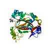 6i9lC 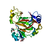 6i9mC 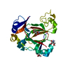 6i9nC  4gjzS S: Starting model for refinement C: citing same article ( |
|---|---|
| Similar structure data |
-Links
 PDBj
PDBj

- Assembly
| Deposited unit | 
| ||||||||
|---|---|---|---|---|---|---|---|---|---|
| 1 |
| ||||||||
| Unit cell |
|
-Components
| #1: Protein | Mass: 29733.547 Da / Num. of mol.: 1 Source method: isolated from a genetically manipulated source Source: (gene. exp.) Homo sapiens (human) / Gene: KDM8, JMJD5 / Production host:  References: UniProt: Q8N371, [protein]-arginine 3-hydroxylase, Hydrolases; Acting on peptide bonds (peptidases) | ||||
|---|---|---|---|---|---|
| #2: Chemical | ChemComp-MN /   Mass: 54.938 Da / Num. of mol.: 1 / Source method: obtained synthetically / Formula: Mn Mass: 54.938 Da / Num. of mol.: 1 / Source method: obtained synthetically / Formula: Mn | ||||
| #3: Chemical | ChemComp-O2U / [(  Mass: 173.123 Da / Num. of mol.: 1 / Source method: obtained synthetically / Formula: C6H7NO5 / Feature type: SUBJECT OF INVESTIGATION Mass: 173.123 Da / Num. of mol.: 1 / Source method: obtained synthetically / Formula: C6H7NO5 / Feature type: SUBJECT OF INVESTIGATION | ||||
| #4: Chemical |   Mass: 92.094 Da / Num. of mol.: 2 / Source method: obtained synthetically / Formula: C3H8O3 Mass: 92.094 Da / Num. of mol.: 2 / Source method: obtained synthetically / Formula: C3H8O3#5: Water | ChemComp-HOH / |  Mass: 18.015 Da / Num. of mol.: 290 / Source method: isolated from a natural source / Formula: H2O Mass: 18.015 Da / Num. of mol.: 290 / Source method: isolated from a natural source / Formula: H2OHas ligand of interest | Y | |
-Experimental details
-Experiment
| Experiment | Method: X-RAY DIFFRACTION / Number of used crystals: 1 |
|---|
- Sample preparation
| Crystal | Density Matthews: 2.11 Å3/Da / Density % sol: 41.58 % |
|---|---|
| Crystal grow | Temperature: 298 K / Method: vapor diffusion, sitting drop / pH: 6.5 Details: Sample: 22 mg/mL JMJD5, 1.5 mM MnCl2, 5 mM compound (IS-52); Reservoir: 0.1 M Bis-Tris pH 6.5, 15.0 % PEG3350, 0.002 M MnCl2; Cryo-protection: 25% (v/v) glycerol; Method: 300 nL sitting ...Details: Sample: 22 mg/mL JMJD5, 1.5 mM MnCl2, 5 mM compound (IS-52); Reservoir: 0.1 M Bis-Tris pH 6.5, 15.0 % PEG3350, 0.002 M MnCl2; Cryo-protection: 25% (v/v) glycerol; Method: 300 nL sitting drops (sample:well, 2:1 ratio) |
-Data collection
| Diffraction | Mean temperature: 100 K / Serial crystal experiment: N | ||||||||||||||||||||||||
|---|---|---|---|---|---|---|---|---|---|---|---|---|---|---|---|---|---|---|---|---|---|---|---|---|---|
| Diffraction source | Source: SYNCHROTRON / Site: Diamond  / Beamline: I02 / Wavelength: 0.98 Å / Beamline: I02 / Wavelength: 0.98 Å | ||||||||||||||||||||||||
| Detector | Type: DECTRIS PILATUS 6M / Detector: PIXEL / Date: May 5, 2015 / Details: MIRRORS | ||||||||||||||||||||||||
| Radiation | Monochromator: SI 111 / Protocol: SINGLE WAVELENGTH / Monochromatic (M) / Laue (L): M / Scattering type: x-ray | ||||||||||||||||||||||||
| Radiation wavelength | Wavelength: 0.98 Å / Relative weight: 1 | ||||||||||||||||||||||||
| Reflection | Resolution: 1.49→78.23 Å / Num. obs: 41815 / % possible obs: 100 % / Redundancy: 7 % / Biso Wilson estimate: 14 Å2 / CC1/2: 0.996 / Rpim(I) all: 0.04 / Rrim(I) all: 0.107 / Rsym value: 0.099 / Net I/σ(I): 11 | ||||||||||||||||||||||||
| Reflection shell | Diffraction-ID: 1
|
- Processing
| Software |
| |||||||||||||||||||||||||||||||||||||||||||||||||||||||
|---|---|---|---|---|---|---|---|---|---|---|---|---|---|---|---|---|---|---|---|---|---|---|---|---|---|---|---|---|---|---|---|---|---|---|---|---|---|---|---|---|---|---|---|---|---|---|---|---|---|---|---|---|---|---|---|---|
| Refinement | Method to determine structure: MOLECULAR REPLACEMENT Starting model: 4GJZ Resolution: 1.49→49.853 Å / SU ML: 0.12 / Cross valid method: THROUGHOUT / σ(F): 1.35 / Phase error: 14.13 / Stereochemistry target values: ML
| |||||||||||||||||||||||||||||||||||||||||||||||||||||||
| Solvent computation | Shrinkage radii: 0.9 Å / VDW probe radii: 1.11 Å / Solvent model: FLAT BULK SOLVENT MODEL | |||||||||||||||||||||||||||||||||||||||||||||||||||||||
| Displacement parameters | Biso max: 70.15 Å2 / Biso mean: 21.1625 Å2 / Biso min: 6.89 Å2 | |||||||||||||||||||||||||||||||||||||||||||||||||||||||
| Refinement step | Cycle: final / Resolution: 1.49→49.853 Å
| |||||||||||||||||||||||||||||||||||||||||||||||||||||||
| LS refinement shell | Refine-ID: X-RAY DIFFRACTION / Rfactor Rfree error: 0 / % reflection obs: 100 %
|
