 Movie
Movie Controller


+ Open data

- Basic information
| Entry | Database: PDB / ID: 7uoa | ||||||
|---|---|---|---|---|---|---|---|
| Title | MAGEA4-MTP1 linear peptide complex | ||||||
 Components Components |
| ||||||
 Keywords Keywords | PROTEIN BINDING / Mage homology domain MHD | ||||||
| Function / homology |  Function and homology information Function and homology informationMelanoma associated antigen, N-terminal / Melanoma associated antigen family N terminal / Melanoma associated antigen family N terminal / MAGE conserved domain profile. / MAGE homology domain / Melanoma-associated antigen / MAGE homology domain, winged helix WH1 motif / MAGE homology domain, winged helix WH2 motif / MAGE homology domain / Melanoma-associated antigen Similarity search - Domain/homology | ||||||
| Biological species |  Homo sapiens (human) Homo sapiens (human)synthetic construct (others) | ||||||
| Method |  X-RAY DIFFRACTION / SYNCHROTRON / MOLECULAR REPLACEMENT / Resolution: 3.5 Å X-RAY DIFFRACTION / SYNCHROTRON / MOLECULAR REPLACEMENT / Resolution: 3.5 Å | ||||||
 Authors Authors | Williams, R.S. / Tumbale, P.S. | ||||||
| Funding support |  United States, 1items United States, 1items
| ||||||
 Citation Citation | Journal: J.Med.Chem. / Year: 2022 Title: Discovery and Structural Basis of the Selectivity of Potent Cyclic Peptide Inhibitors of MAGE-A4. Authors: Fleming, M.C. / Chiou, L.F. / Tumbale, P.P. / Droby, G.N. / Lim, J. / Norris-Drouin, J.L. / Williams, J.G. / Pearce, K.H. / Williams, R.S. / Vaziri, C. / Bowers, A.A. | ||||||
| History |
|
- Structure visualization
| Structure viewer | Molecule: MolmilJmol/JSmol |
|---|
- Downloads & links
-Download
| PDBx/mmCIF format | 7uoa.cif.gz | 102.5 KB | Display | PDBx/mmCIF format |
|---|---|---|---|---|
| PDB format | pdb7uoa.ent.gz | 77.2 KB | Display | PDB format |
| PDBx/mmJSON format | 7uoa.json.gz | Tree view | PDBx/mmJSON format | |
| Others |  Other downloads Other downloads |
-Validation report
| Arichive directory | https://data.pdbj.org/pub/pdb/validation_reports/uo/7uoaftp://data.pdbj.org/pub/pdb/validation_reports/uo/7uoa | HTTPS FTP |
|---|
-Related structure data
| Related structure data | 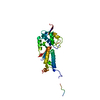 2wa0S S: Starting model for refinement |
|---|---|
| Similar structure data |
-Links
 PDBj
PDBj- Assembly
| Deposited unit | 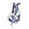
| ||||||||
|---|---|---|---|---|---|---|---|---|---|
| 1 |
| ||||||||
| Unit cell |
|
-Components
| #1: Protein | Mass: 24997.490 Da / Num. of mol.: 1 / Fragment: UNP residues 101-317 Source method: isolated from a genetically manipulated source Source: (gene. exp.) Homo sapiens (human) / Gene: MAGEA4 / Production host:  |
|---|---|
| #2: Protein/peptide | Mass: 1361.526 Da / Num. of mol.: 1 / Source method: obtained synthetically / Source: (synth.) synthetic construct (others) |
| #3: Chemical | ChemComp-EDO /   Mass: 62.068 Da / Num. of mol.: 1 / Source method: obtained synthetically / Formula: C2H6O2 Mass: 62.068 Da / Num. of mol.: 1 / Source method: obtained synthetically / Formula: C2H6O2 |
| #4: Chemical | ChemComp-TRS /   Mass: 122.143 Da / Num. of mol.: 1 / Source method: obtained synthetically / Formula: C4H12NO3 / Comment: pH buffer*YM Mass: 122.143 Da / Num. of mol.: 1 / Source method: obtained synthetically / Formula: C4H12NO3 / Comment: pH buffer*YM |
| Has ligand of interest | N |
-Experimental details
-Experiment
| Experiment | Method: X-RAY DIFFRACTION / Number of used crystals: 1 |
|---|
- Sample preparation
| Crystal | Density Matthews: 3.71 Å3/Da / Density % sol: 66.85 % |
|---|---|
| Crystal grow | Temperature: 293 K / Method: vapor diffusion, sitting drop / Details: Tris-HCl, magnesium chloride, ethanol |
-Data collection
| Diffraction | Mean temperature: 100 K / Serial crystal experiment: N |
|---|---|
| Diffraction source | Source: SYNCHROTRON / Site: APS / Beamline: 22-ID / Wavelength: 1 Å |
| Detector | Type: DECTRIS EIGER X 16M / Detector: PIXEL / Date: Feb 4, 2021 |
| Radiation | Protocol: SINGLE WAVELENGTH / Monochromatic (M) / Laue (L): M / Scattering type: x-ray |
| Radiation wavelength | Wavelength: 1 Å / Relative weight: 1 |
| Reflection | Resolution: 3.5→50 Å / Num. obs: 5488 / % possible obs: 98.24 % / Redundancy: 10.5 % / CC1/2: 0.992 / Net I/σ(I): 8.65 |
| Reflection shell | Resolution: 3.5→3.626 Å / Num. unique obs: 5248 / CC1/2: 0.967 |
- Processing
| Software |
| ||||||||||||||||||||||||||||||||||||||||||||||||||||||||
|---|---|---|---|---|---|---|---|---|---|---|---|---|---|---|---|---|---|---|---|---|---|---|---|---|---|---|---|---|---|---|---|---|---|---|---|---|---|---|---|---|---|---|---|---|---|---|---|---|---|---|---|---|---|---|---|---|---|
| Refinement | Method to determine structure: MOLECULAR REPLACEMENT Starting model: PDB entry 2WA0 Resolution: 3.5→49.61 Å / SU ML: 0.5 / Cross valid method: THROUGHOUT / σ(F): 1.41 / Phase error: 32.17 / Stereochemistry target values: ML
| ||||||||||||||||||||||||||||||||||||||||||||||||||||||||
| Solvent computation | Shrinkage radii: 0.9 Å / VDW probe radii: 1.11 Å / Solvent model: FLAT BULK SOLVENT MODEL | ||||||||||||||||||||||||||||||||||||||||||||||||||||||||
| Displacement parameters | Biso max: 180.22 Å2 / Biso mean: 116.098 Å2 / Biso min: 70.69 Å2 | ||||||||||||||||||||||||||||||||||||||||||||||||||||||||
| Refinement step | Cycle: final / Resolution: 3.5→49.61 Å
| ||||||||||||||||||||||||||||||||||||||||||||||||||||||||
| LS refinement shell | Refine-ID: X-RAY DIFFRACTION / Rfactor Rfree error: 0 / Total num. of bins used: 7
| ||||||||||||||||||||||||||||||||||||||||||||||||||||||||
| Refinement TLS params. | Method: refined / Origin x: -28.3398 Å / Origin y: 14.4404 Å / Origin z: -8.0221 Å
| ||||||||||||||||||||||||||||||||||||||||||||||||||||||||
| Refinement TLS group |
|
