 Movie
Movie Controller

[English] 日本語
 Yorodumi
- PDB-7ukj: In situ cryo-EM structure of bacteriophage Sf6 portal:gp7 complex... -
+ Open data

- Basic information
| Entry | Database: PDB / ID: 7ukj | |||||||||||||||||||||||||||
|---|---|---|---|---|---|---|---|---|---|---|---|---|---|---|---|---|---|---|---|---|---|---|---|---|---|---|---|---|
| Title | In situ cryo-EM structure of bacteriophage Sf6 portal:gp7 complex at 2.7A resolution | |||||||||||||||||||||||||||
 Components Components | Gene 14 protein | |||||||||||||||||||||||||||
 Keywords Keywords | VIRAL PROTEIN / in situ / phage / portal / gp7 / gp3 / structural protein | |||||||||||||||||||||||||||
| Function / homology |  Function and homology information Function and homology informationendo-1,3-alpha-L-rhamnosidase activity / symbiont entry into host cell via disruption of host cell envelope lipopolysaccharide / virus tail, fiber / symbiont entry into host cell via disruption of host cell envelope / virion attachment to host cell Similarity search - Function | |||||||||||||||||||||||||||
| Biological species |  Shigella phage Sf6 (virus) Shigella phage Sf6 (virus) | |||||||||||||||||||||||||||
| Method | ELECTRON MICROSCOPY / single particle reconstruction / cryo EM / Resolution: 3.6 Å | |||||||||||||||||||||||||||
 Authors Authors | Li, F. / Cingolani, G. / Hou, C. | |||||||||||||||||||||||||||
| Funding support |  United States, 1items United States, 1items
| |||||||||||||||||||||||||||
 Citation Citation | Journal: Sci Adv / Year: 2022 Title: High-resolution cryo-EM structure of the virus Sf6 genome delivery tail machine. Authors: Fenglin Li / Chun-Feng David Hou / Ruoyu Yang / Richard Whitehead / Carolyn M Teschke / Gino Cingolani / Abstract: Sf6 is a bacterial virus that infects the human pathogen Here, we describe the cryo-electron microscopy structure of the Sf6 tail machine before DNA ejection, which we determined at a 2.7-angstrom ...Sf6 is a bacterial virus that infects the human pathogen Here, we describe the cryo-electron microscopy structure of the Sf6 tail machine before DNA ejection, which we determined at a 2.7-angstrom resolution. We built de novo structures of all tail components and resolved four symmetry-mismatched interfaces. Unexpectedly, we found that the tail exists in two conformations, rotated by ~6° with respect to the capsid. The two tail conformers are identical in structure but differ solely in how the portal and head-to-tail adaptor carboxyl termini bond with the capsid at the fivefold vertex, similar to a diamond held over a five-pronged ring in two nonidentical states. Thus, in the mature Sf6 tail, the portal structure does not morph locally to accommodate the symmetry mismatch but exists in two energetic minima rotated by a discrete angle. We propose that the design principles of the Sf6 tail are conserved across P22-like Podoviridae. | |||||||||||||||||||||||||||
| History |
|
- Structure visualization
| Structure viewer | Molecule: MolmilJmol/JSmol |
|---|
- Downloads & links
-Download
| PDBx/mmCIF format | 7ukj.cif.gz | 312.8 KB | Display | PDBx/mmCIF format |
|---|---|---|---|---|
| PDB format | pdb7ukj.ent.gz | 258 KB | Display | PDB format |
| PDBx/mmJSON format | 7ukj.json.gz | Tree view | PDBx/mmJSON format | |
| Others |  Other downloads Other downloads |
-Validation report
| Arichive directory | https://data.pdbj.org/pub/pdb/validation_reports/uk/7ukjftp://data.pdbj.org/pub/pdb/validation_reports/uk/7ukj | HTTPS FTP |
|---|
-Related structure data
| Related structure data |  26582MC 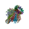 7sfsC  7sg7C  7sp4C  7spuC M: map data used to model this data C: citing same article ( |
|---|---|
| Similar structure data |


-Links
 PDBj
PDBj- Assembly
| Deposited unit | 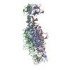
|
|---|---|
| 1 |
|
-Components
| #1: Protein | Mass: 67118.141 Da / Num. of mol.: 3 / Fragment: Tailspike / Source method: isolated from a natural source / Source: (natural) Shigella phage Sf6 (virus) / References: UniProt: Q716G1Has ligand of interest | N | Has protein modification | N | |
|---|
-Experimental details
-Experiment
| Experiment | Method: ELECTRON MICROSCOPY |
|---|---|
| EM experiment | Aggregation state: PARTICLE / 3D reconstruction method: single particle reconstruction |
- Sample preparation
| Component | Name: Shigella virus Sf6 / Type: VIRUS / Entity ID: all / Source: NATURAL |
|---|---|
| Source (natural) | Organism: Shigella virus Sf6 |
| Details of virus | Empty: YES / Enveloped: YES / Isolate: OTHER / Type: VIRION |
| Buffer solution | pH: 7.5 |
| Specimen | Embedding applied: NO / Shadowing applied: NO / Staining applied: NO / Vitrification applied: YES |
| Vitrification | Cryogen name: ETHANE |
- Electron microscopy imaging
| Experimental equipment |  Model: Titan Krios / Image courtesy: FEI Company |
|---|---|
| Microscopy | Model: TFS KRIOS |
| Electron gun | Electron source:  FIELD EMISSION GUN / Accelerating voltage: 300 kV / Illumination mode: OTHER FIELD EMISSION GUN / Accelerating voltage: 300 kV / Illumination mode: OTHER |
| Electron lens | Mode: OTHER / Nominal magnification: 81000 X / Nominal defocus max: 1500 nm / Nominal defocus min: 500 nm / Cs: 2.7 mm / C2 aperture diameter: 100 µm |
| Specimen holder | Cryogen: NITROGEN |
| Image recording | Electron dose: 50 e/Å2 / Film or detector model: GATAN K3 (6k x 4k) / Num. of real images: 7977 |
- Processing
| Software | Name: PHENIX / Version: 1.19.2_4158: / Classification: refinement | ||||||||||||||||||||||||
|---|---|---|---|---|---|---|---|---|---|---|---|---|---|---|---|---|---|---|---|---|---|---|---|---|---|
| EM software |
| ||||||||||||||||||||||||
| CTF correction | Type: PHASE FLIPPING AND AMPLITUDE CORRECTION | ||||||||||||||||||||||||
| Particle selection | Num. of particles selected: 67439 | ||||||||||||||||||||||||
| 3D reconstruction | Resolution: 3.6 Å / Resolution method: FSC 0.143 CUT-OFF / Num. of particles: 94000 / Symmetry type: POINT | ||||||||||||||||||||||||
| Refine LS restraints |
|
