 Movie
Movie Controller

[English] 日本語
 Yorodumi
- EMDB-25101: In situ cryo-EM structure of bacteriophage Sf6 portal:gp7 complex... -
+ Open data

- Basic information
| Entry |  | |||||||||
|---|---|---|---|---|---|---|---|---|---|---|
| Title | In situ cryo-EM structure of bacteriophage Sf6 portal:gp7 complex at 2.7A resolution | |||||||||
 Map data Map data | ||||||||||
 Sample Sample |
| |||||||||
 Keywords Keywords | in situ / phage / portal / gp7 / gp3 / STRUCTURAL PROTEIN | |||||||||
| Function / homology | Tail accessory factor GP4 / Peptidoglycan hydrolase Gp4 superfamily / P22 tail accessory factor / Phage P22-like portal protein / Phage P22-like portal protein / symbiont genome ejection through host cell envelope, short tail mechanism / metal ion binding / Gene 7 protein / Gene 3 protein Function and homology information Function and homology information | |||||||||
| Biological species |  Shigella phage Sf6 (virus) / Shigella virus Sf6 Shigella phage Sf6 (virus) / Shigella virus Sf6 | |||||||||
| Method | single particle reconstruction / cryo EM / Resolution: 2.76 Å | |||||||||
 Authors Authors | Li F / Cingolani G | |||||||||
| Funding support |  United States, 1 items United States, 1 items
| |||||||||
 Citation Citation | Journal: Sci Adv / Year: 2022 Title: High-resolution cryo-EM structure of the virus Sf6 genome delivery tail machine. Authors: Fenglin Li / Chun-Feng David Hou / Ruoyu Yang / Richard Whitehead / Carolyn M Teschke / Gino Cingolani / Abstract: Sf6 is a bacterial virus that infects the human pathogen Here, we describe the cryo-electron microscopy structure of the Sf6 tail machine before DNA ejection, which we determined at a 2.7-angstrom ...Sf6 is a bacterial virus that infects the human pathogen Here, we describe the cryo-electron microscopy structure of the Sf6 tail machine before DNA ejection, which we determined at a 2.7-angstrom resolution. We built de novo structures of all tail components and resolved four symmetry-mismatched interfaces. Unexpectedly, we found that the tail exists in two conformations, rotated by ~6° with respect to the capsid. The two tail conformers are identical in structure but differ solely in how the portal and head-to-tail adaptor carboxyl termini bond with the capsid at the fivefold vertex, similar to a diamond held over a five-pronged ring in two nonidentical states. Thus, in the mature Sf6 tail, the portal structure does not morph locally to accommodate the symmetry mismatch but exists in two energetic minima rotated by a discrete angle. We propose that the design principles of the Sf6 tail are conserved across P22-like Podoviridae. | |||||||||
| History |
|
- Structure visualization
| Supplemental images |
|---|

- Downloads & links
-EMDB archive
| Map data | emd_25101.map.gz | 393.6 MB | EMDB map data format | |
|---|---|---|---|---|
| Header (meta data) | emd-25101-v30.xmlemd-25101.xml | 11.4 KB 11.4 KB | Display Display | EMDB header |
| FSC (resolution estimation) | emd_25101_fsc.xml | 18 KB | Display | FSC data file |
| Images | 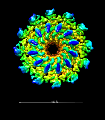 emd_25101.png emd_25101.png | 104.7 KB | ||
| Filedesc metadata | emd-25101.cif.gz | 5.5 KB | ||
| Archive directory |  http://ftp.pdbj.org/pub/emdb/structures/EMD-25101ftp://ftp.pdbj.org/pub/emdb/structures/EMD-25101 http://ftp.pdbj.org/pub/emdb/structures/EMD-25101ftp://ftp.pdbj.org/pub/emdb/structures/EMD-25101 | HTTPS FTP |
-Related structure data
| Related structure data | 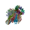 7sfsMC  7sg7C  7sp4C  7spuC  7ukjC M: atomic model generated by this map C: citing same article ( |
|---|---|
| Similar structure data |


-Links
| EMDB pages | EMDB (EBI/PDBe) / EMDataResource |
|---|
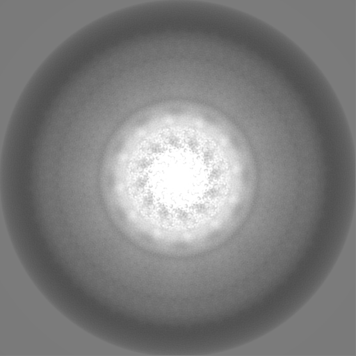
-Map
| File | Download / File: emd_25101.map.gz / Format: CCP4 / Size: 512 MB / Type: IMAGE STORED AS FLOATING POINT NUMBER (4 BYTES) | ||||||||||||||||||||||||||||||||||||
|---|---|---|---|---|---|---|---|---|---|---|---|---|---|---|---|---|---|---|---|---|---|---|---|---|---|---|---|---|---|---|---|---|---|---|---|---|---|
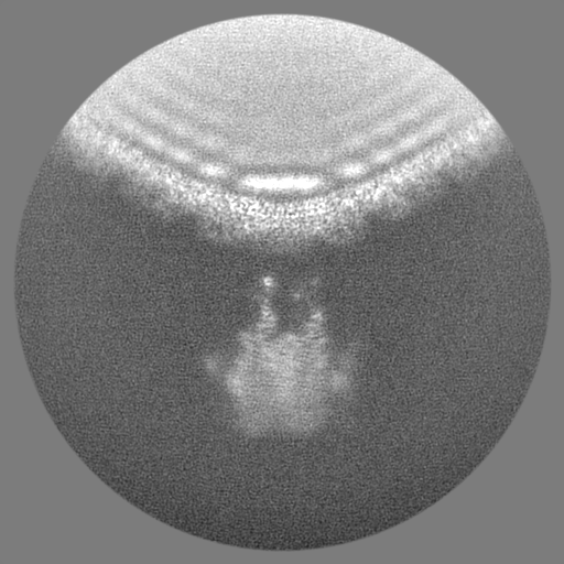
| Projections & slices | Image control
Images are generated by Spider. | ||||||||||||||||||||||||||||||||||||
| Voxel size | X=Y=Z: 1.122 Å | ||||||||||||||||||||||||||||||||||||
| Density |
| ||||||||||||||||||||||||||||||||||||
| Symmetry | Space group: 1 | ||||||||||||||||||||||||||||||||||||
| Details | EMDB XML:
|
 Z (Sec.)
 Y (Row.)
 X (Col.)


-Supplemental data
- Sample components
-Entire : Shigella virus Sf6
| Entire | Name: Shigella virus Sf6 |
|---|---|
| Components |
|
-Supramolecule #1: Shigella virus Sf6
| Supramolecule | Name: Shigella virus Sf6 / type: virus / ID: 1 / Parent: 0 / Macromolecule list: all / NCBI-ID: 10761 / Sci species name: Shigella virus Sf6 / Virus type: VIRION / Virus isolate: OTHER / Virus enveloped: Yes / Virus empty: Yes |
|---|
-Macromolecule #1: Gene 3 protein
| Macromolecule | Name: Gene 3 protein / type: protein_or_peptide / ID: 1 / Number of copies: 12 / Enantiomer: LEVO |
|---|---|
| Source (natural) | Organism: Shigella phage Sf6 (virus) |
| Molecular weight | Theoretical: 79.558227 KDa |
| Sequence | String: MAETLEKKHE RIMLRFDRAY SPQKEVREKC IEATRFARVP GGQWEGATAA GTKLDEQFEK YPKFEINKVA TELNRIIAEY RNNRITVKF RPGDREASEE LANKLNGLFR ADYEETDGGE ACDNAFDDAA TGGFGCFRLT SMLVNEYDPM DDRQRIAIEP I YDPSRSVW ...String: MAETLEKKHE RIMLRFDRAY SPQKEVREKC IEATRFARVP GGQWEGATAA GTKLDEQFEK YPKFEINKVA TELNRIIAEY RNNRITVKF RPGDREASEE LANKLNGLFR ADYEETDGGE ACDNAFDDAA TGGFGCFRLT SMLVNEYDPM DDRQRIAIEP I YDPSRSVW FDPDAKKYDK SDALWAFCMY SLSPEKYEAE YGKKPPTSLD VTSMTSWEYN WFGADVIYIA KYYEVRKESV DV ISYRHPI TGEIATYDSD QVEDIEDELA IAGFHEVARR SVKRRRVYVS VVDGDGFLEK PRRIPGEHIP LIPVYGKRWF IDD IERVEG HIAKAMDPQR LYNLQVSMLA DTAAQDPGQI PIVGMEQIRG LEKHWEARNK KRPAFLPLRE VRDKSGNIIA GATP AGYTQ PAVMNQALAA LLQQTSADIQ EVTGGSQAMQ QMPSNIAQET VNNLMNRADM ASFIYLDNMA KSLKRAGEVW LSMAR EVYG SEREVRIVNE DGSDDIAVLS AQVVDRQTGA VVALNDLSVG RYDVTVDVGP SYTARRDATV SVLTNVLSSM LPTDPM RPA IQGIILDNID GEGLDDFKEY NRNQLLISGI AKPRNEKEQQ IVQQAQMAAQ SQPNPEMVLA QAQMVAAQAE AQKATNE TA QTQIKAFTAQ QDAMESQANT VYKLAQARNI DDKAVMEAIR LLKDVAESQQ QQFQSPPQSP ADLMPS UniProtKB: Gene 3 protein |
-Macromolecule #2: Gene 7 protein
| Macromolecule | Name: Gene 7 protein / type: protein_or_peptide / ID: 2 / Number of copies: 12 / Enantiomer: LEVO |
|---|---|
| Source (natural) | Organism: Shigella phage Sf6 (virus) |
| Molecular weight | Theoretical: 17.753889 KDa |
| Sequence | String: MATVLTKGEI VLFALRKFAI ASNASLTDVE PQSIEDGVND LEDMMSEWMI NPGDIGYAFA TGDEQPLPDD ESGLPRKYKH AVGYQLLLR MLSDYSLEPT PQVLSNAQRS YDALMTDTLV VPSMRRRGDF PVGQGNKYDV FTSDRYYPGD LPLIDGDIPN A UniProtKB: Gene 7 protein |
-Experimental details
-Structure determination
| Method | cryo EM |
|---|---|
 Processing Processing | single particle reconstruction |
| Aggregation state | particle |
-Sample preparation
| Buffer | pH: 7.5 |
|---|---|
| Grid | Model: Quantifoil R2/1 / Material: COPPER / Mesh: 300 |
| Vitrification | Cryogen name: ETHANE |
- Electron microscopy
| Microscope | TFS KRIOS |
|---|---|
| Image recording | Film or detector model: GATAN K3 (6k x 4k) / Number real images: 7977 / Average electron dose: 50.0 e/Å2 |
| Electron beam | Acceleration voltage: 300 kV / Electron source:  FIELD EMISSION GUN FIELD EMISSION GUN |
| Electron optics | C2 aperture diameter: 100.0 µm / Illumination mode: OTHER / Imaging mode: OTHER / Cs: 2.7 mm / Nominal defocus max: 1.5 µm / Nominal defocus min: 0.5 µm / Nominal magnification: 81000 |
| Sample stage | Cooling holder cryogen: NITROGEN |
| Experimental equipment |  Model: Titan Krios / Image courtesy: FEI Company |

