 Movie
Movie Controller

[English] 日本語
 Yorodumi
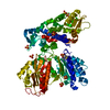
- PDB-7uch: AprA Methyltransferase 1 - GNAT in complex with Mn2+ , SAM, and D... -
+ Open data

- Basic information
| Entry | Database: PDB / ID: 7uch | ||||||||||||||||||
|---|---|---|---|---|---|---|---|---|---|---|---|---|---|---|---|---|---|---|---|
| Title | AprA Methyltransferase 1 - GNAT in complex with Mn2+ , SAM, and Di-methyl-malonate | ||||||||||||||||||
 Components Components | AprA Methyltransferase 1 | ||||||||||||||||||
 Keywords Keywords | TRANSFERASE / polyketide synthase | ||||||||||||||||||
| Function / homology |  Function and homology information Function and homology information | ||||||||||||||||||
| Biological species |  Moorena bouillonii (bacteria) Moorena bouillonii (bacteria) | ||||||||||||||||||
| Method |  X-RAY DIFFRACTION / SYNCHROTRON / MOLECULAR REPLACEMENT / Resolution: 2.18 Å X-RAY DIFFRACTION / SYNCHROTRON / MOLECULAR REPLACEMENT / Resolution: 2.18 Å | ||||||||||||||||||
 Authors Authors | Skiba, M.A. / Lao, Y. / Smith, J.L. | ||||||||||||||||||
| Funding support |  United States, 5items United States, 5items
| ||||||||||||||||||
 Citation Citation | Journal: Acs Chem.Biol. / Year: 2022 Title: Structural Basis for Control of Methylation Extent in Polyketide Synthase Metal-Dependent C -Methyltransferases. Authors: Lao, Y. / Skiba, M.A. / Chun, S.W. / Narayan, A.R.H. / Smith, J.L. #1: Journal: ACS Chem Biol / Year: 2018Title: Biosynthesis of t-Butyl in Apratoxin A: Functional Analysis and Architecture of a PKS Loading Module. Authors: Meredith A Skiba / Andrew P Sikkema / Nathan A Moss / Andrew N Lowell / Min Su / Rebecca M Sturgis / Lena Gerwick / William H Gerwick / David H Sherman / Janet L Smith / Abstract: The unusual feature of a t-butyl group is found in several marine-derived natural products including apratoxin A, a Sec61 inhibitor produced by the cyanobacterium Moorea bouillonii PNG 5-198. Here, ...The unusual feature of a t-butyl group is found in several marine-derived natural products including apratoxin A, a Sec61 inhibitor produced by the cyanobacterium Moorea bouillonii PNG 5-198. Here, we determine that the apratoxin A t-butyl group is formed as a pivaloyl acyl carrier protein (ACP) by AprA, the polyketide synthase (PKS) loading module of the apratoxin A biosynthetic pathway. AprA contains an inactive "pseudo" GCN5-related N-acetyltransferase domain (ΨGNAT) flanked by two methyltransferase domains (MT1 and MT2) that differ distinctly in sequence. Structural, biochemical, and precursor incorporation studies reveal that MT2 catalyzes unusually coupled decarboxylation and methylation reactions to transform dimethylmalonyl-ACP, the product of MT1, to pivaloyl-ACP. Further, pivaloyl-ACP synthesis is primed by the fatty acid synthase malonyl acyltransferase (FabD), which compensates for the ΨGNAT and provides the initial acyl-transfer step to form AprA malonyl-ACP. Additionally, images of AprA from negative stain electron microscopy reveal multiple conformations that may facilitate the individual catalytic steps of the multienzyme module. | ||||||||||||||||||
| History |
|
- Structure visualization
| Structure viewer | Molecule: MolmilJmol/JSmol |
|---|
- Downloads & links
-Download
| PDBx/mmCIF format | 7uch.cif.gz | 188.5 KB | Display | PDBx/mmCIF format |
|---|---|---|---|---|
| PDB format | pdb7uch.ent.gz | 115.8 KB | Display | PDB format |
| PDBx/mmJSON format | 7uch.json.gz | Tree view | PDBx/mmJSON format | |
| Others |  Other downloads Other downloads |
-Validation report
| Arichive directory | https://data.pdbj.org/pub/pdb/validation_reports/uc/7uchftp://data.pdbj.org/pub/pdb/validation_reports/uc/7uch | HTTPS FTP |
|---|
-Related structure data
| Related structure data |  7uciC  7uclC  6b3aS S: Starting model for refinement C: citing same article ( |
|---|---|
| Similar structure data |

-Links
 PDBj
PDBj


- Assembly
| Deposited unit | 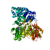
| ||||||||||||
|---|---|---|---|---|---|---|---|---|---|---|---|---|---|
| 1 |
| ||||||||||||
| Unit cell |
|
-Components
-Protein , 1 types, 1 molecules A
| #1: Protein | Mass: 74825.836 Da / Num. of mol.: 1 Source method: isolated from a genetically manipulated source Source: (gene. exp.) Moorena bouillonii (bacteria) / Production host: |
|---|
-Non-polymers , 5 types, 323 molecules 


| #2: Chemical | ChemComp-MN /  Mass: 54.938 Da / Num. of mol.: 1 / Source method: obtained synthetically / Formula: Mn / Feature type: SUBJECT OF INVESTIGATION Mass: 54.938 Da / Num. of mol.: 1 / Source method: obtained synthetically / Formula: Mn / Feature type: SUBJECT OF INVESTIGATION |
|---|---|
| #3: Chemical | ChemComp-GOL /  Mass: 92.094 Da / Num. of mol.: 1 / Source method: obtained synthetically / Formula: C3H8O3 Mass: 92.094 Da / Num. of mol.: 1 / Source method: obtained synthetically / Formula: C3H8O3 |
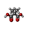
| #4: Chemical | ChemComp-MU6 /  Mass: 132.115 Da / Num. of mol.: 1 / Source method: obtained synthetically / Formula: C5H8O4 Mass: 132.115 Da / Num. of mol.: 1 / Source method: obtained synthetically / Formula: C5H8O4 |
| #5: Chemical | ChemComp-SAH /  Type: L-peptide linking / Mass: 384.411 Da / Num. of mol.: 1 / Source method: obtained synthetically / Formula: C14H20N6O5S / Feature type: SUBJECT OF INVESTIGATION Type: L-peptide linking / Mass: 384.411 Da / Num. of mol.: 1 / Source method: obtained synthetically / Formula: C14H20N6O5S / Feature type: SUBJECT OF INVESTIGATION |
| #6: Water | ChemComp-HOH / Mass: 18.015 Da / Num. of mol.: 319 / Source method: isolated from a natural source / Formula: H2O |
-Details
| Has ligand of interest | Y |
|---|
-Experimental details
-Experiment
| Experiment | Method: X-RAY DIFFRACTION / Number of used crystals: 1 |
|---|
- Sample preparation
| Crystal | Density Matthews: 2.43 Å3/Da / Density % sol: 49.37 % |
|---|---|
| Crystal grow | Temperature: 293 K / Method: vapor diffusion, sitting drop Details: 11mg/mL protein in 50mM Tris pH7.4, 50mM NaCl, 10%(V/V) glycerol, 1mM S-adenosyl-L-homocysteine (SAH), and 5mM MnCl2. Well solution: 15% PEG 3350, 0.1M di-methyl-malonate Protein to well solution ratio is 2:2 |
-Data collection
| Diffraction | Mean temperature: 100 K / Serial crystal experiment: N |
|---|---|
| Diffraction source | Source: SYNCHROTRON / Site: APS / Beamline: 23-ID-B / Wavelength: 1.033 Å |
| Detector | Type: DECTRIS EIGER X 16M / Detector: PIXEL / Date: Nov 9, 2017 |
| Radiation | Monochromator: M / Protocol: SINGLE WAVELENGTH / Monochromatic (M) / Laue (L): M / Scattering type: x-ray |
| Radiation wavelength | Wavelength: 1.033 Å / Relative weight: 1 |
| Reflection | Resolution: 2.18→45.23 Å / Num. obs: 38749 / % possible obs: 99.87 % / Redundancy: 10.6 % / Biso Wilson estimate: 33.02 Å2 / CC1/2: 0.995 / CC star: 0.999 / Net I/σ(I): 7.37 |
| Reflection shell | Resolution: 2.181→2.259 Å / Mean I/σ(I) obs: 1.18 / Num. unique obs: 3805 / CC1/2: 0.495 / CC star: 0.814 / % possible all: 99.42 |
- Processing
| Software |
| ||||||||||||||||||||||||
|---|---|---|---|---|---|---|---|---|---|---|---|---|---|---|---|---|---|---|---|---|---|---|---|---|---|
| Refinement | Method to determine structure: MOLECULAR REPLACEMENT Starting model: 6B3A Resolution: 2.18→45.23 Å / SU ML: 0.3282 / Cross valid method: FREE R-VALUE / σ(F): 1.35 / Phase error: 26.5831 Stereochemistry target values: GeoStd + Monomer Library + CDL v1.2
| ||||||||||||||||||||||||
| Solvent computation | Shrinkage radii: 0.9 Å / VDW probe radii: 1.1 Å / Solvent model: FLAT BULK SOLVENT MODEL | ||||||||||||||||||||||||
| Displacement parameters | Biso mean: 40.78 Å2 | ||||||||||||||||||||||||
| Refinement step | Cycle: LAST / Resolution: 2.18→45.23 Å
| ||||||||||||||||||||||||
| Refine LS restraints |
| ||||||||||||||||||||||||
| LS refinement shell | Resolution: 2.18→2.21 Å
|
