 Movie
Movie Controller

[English] 日本語
 Yorodumi
- PDB-7uci: SxtA Methyltransferase and decarboxylase didomain in complex with... -
+ Open data

- Basic information
| Entry | Database: PDB / ID: 7uci | ||||||||||||
|---|---|---|---|---|---|---|---|---|---|---|---|---|---|
| Title | SxtA Methyltransferase and decarboxylase didomain in complex with Mn2+ and SAH | ||||||||||||
 Components Components | Polyketide synthase-related protein | ||||||||||||
 Keywords Keywords | TRANSFERASE / Methyltransferase | ||||||||||||
| Function / homology |  Function and homology information Function and homology information8-amino-7-oxononanoate synthase activity / biotin biosynthetic process / phosphopantetheine binding / pyridoxal phosphate binding / metal ion binding Similarity search - Function | ||||||||||||
| Biological species |  Cylindrospermopsis raciborskii (bacteria) Cylindrospermopsis raciborskii (bacteria) | ||||||||||||
| Method |  X-RAY DIFFRACTION / SYNCHROTRON / MOLECULAR REPLACEMENT / Resolution: 2.6 Å X-RAY DIFFRACTION / SYNCHROTRON / MOLECULAR REPLACEMENT / Resolution: 2.6 Å | ||||||||||||
 Authors Authors | Lao, Y. / Skiba, M.A. / Smith, J.L. | ||||||||||||
| Funding support |  United States, 3items United States, 3items
| ||||||||||||
 Citation Citation | Journal: Acs Chem.Biol. / Year: 2022 Title: Structural Basis for Control of Methylation Extent in Polyketide Synthase Metal-Dependent C -Methyltransferases. Authors: Lao, Y. / Skiba, M.A. / Chun, S.W. / Narayan, A.R.H. / Smith, J.L. | ||||||||||||
| History |
|
- Structure visualization
| Structure viewer | Molecule: MolmilJmol/JSmol |
|---|
- Downloads & links
-Download
| PDBx/mmCIF format | 7uci.cif.gz | 631.2 KB | Display | PDBx/mmCIF format |
|---|---|---|---|---|
| PDB format | pdb7uci.ent.gz | 422.4 KB | Display | PDB format |
| PDBx/mmJSON format | 7uci.json.gz | Tree view | PDBx/mmJSON format | |
| Others |  Other downloads Other downloads |
-Validation report
| Arichive directory | https://data.pdbj.org/pub/pdb/validation_reports/uc/7uciftp://data.pdbj.org/pub/pdb/validation_reports/uc/7uci | HTTPS FTP |
|---|
-Related structure data
| Related structure data | 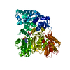 7uchC  7uclC  6b3aS S: Starting model for refinement C: citing same article ( |
|---|---|
| Similar structure data |
-Links
 PDBj
PDBj


- Assembly
| Deposited unit | 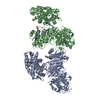
| ||||||||||||
|---|---|---|---|---|---|---|---|---|---|---|---|---|---|
| 1 | 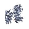
| ||||||||||||
| 2 | 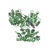
| ||||||||||||
| Unit cell |
|
-Components
| #1: Protein | Mass: 81187.008 Da / Num. of mol.: 2 Source method: isolated from a genetically manipulated source Source: (gene. exp.) Cylindrospermopsis raciborskii (bacteria)Gene: sxtA / Production host: #2: Chemical |   Type: L-peptide linking / Mass: 384.411 Da / Num. of mol.: 2 / Source method: obtained synthetically / Formula: C14H20N6O5S / Feature type: SUBJECT OF INVESTIGATION Type: L-peptide linking / Mass: 384.411 Da / Num. of mol.: 2 / Source method: obtained synthetically / Formula: C14H20N6O5S / Feature type: SUBJECT OF INVESTIGATION#3: Chemical |   Mass: 54.938 Da / Num. of mol.: 2 / Source method: obtained synthetically / Formula: Mn / Feature type: SUBJECT OF INVESTIGATION Mass: 54.938 Da / Num. of mol.: 2 / Source method: obtained synthetically / Formula: Mn / Feature type: SUBJECT OF INVESTIGATION#4: Chemical | ChemComp-GOL / |   Mass: 92.094 Da / Num. of mol.: 1 / Source method: obtained synthetically / Formula: C3H8O3 Mass: 92.094 Da / Num. of mol.: 1 / Source method: obtained synthetically / Formula: C3H8O3#5: Water | ChemComp-HOH / |  Mass: 18.015 Da / Num. of mol.: 87 / Source method: isolated from a natural source / Formula: H2O Mass: 18.015 Da / Num. of mol.: 87 / Source method: isolated from a natural source / Formula: H2OHas ligand of interest | Y | |
|---|
-Experimental details
-Experiment
| Experiment | Method: X-RAY DIFFRACTION / Number of used crystals: 1 |
|---|
- Sample preparation
| Crystal | Density Matthews: 2.37 Å3/Da / Density % sol: 48 % |
|---|---|
| Crystal grow | Temperature: 277 K / Method: vapor diffusion, sitting drop / pH: 6.25 Details: 12-14mg/mL SxtA MT-DC in 50mM Tris pH7.4, 50mM NaCl, 10%(v/v) glycerol, 5mM S-adenosyl-L-methionine (SAM), and 5mM MnCl2. Well solution: 5-8% PEG 20K, and 0.1M MES pH 6.25-6.5 PH range: 6.25-6.5 |
-Data collection
| Diffraction | Mean temperature: 100 K / Serial crystal experiment: N |
|---|---|
| Diffraction source | Source: SYNCHROTRON / Site: APS / Beamline: 23-ID-B / Wavelength: 1.033 Å |
| Detector | Type: DECTRIS EIGER X 16M / Detector: PIXEL / Date: Feb 12, 2019 |
| Radiation | Monochromator: M / Protocol: SINGLE WAVELENGTH / Monochromatic (M) / Laue (L): M / Scattering type: x-ray |
| Radiation wavelength | Wavelength: 1.033 Å / Relative weight: 1 |
| Reflection | Resolution: 2.6→42.43 Å / Num. obs: 44788 / % possible obs: 97.8 % / Redundancy: 3.6 % / Biso Wilson estimate: 51.19 Å2 / CC1/2: 0.995 / CC star: 0.999 / Rmerge(I) obs: 0.1261 / Net I/σ(I): 8.57 |
| Reflection shell | Resolution: 2.6→2.69 Å / Redundancy: 3.5 % / Mean I/σ(I) obs: 1.39 / Num. unique obs: 4306 / CC1/2: 0.696 / CC star: 0.906 / % possible all: 94.19 |
- Processing
| Software |
| ||||||||||||||||||||||||
|---|---|---|---|---|---|---|---|---|---|---|---|---|---|---|---|---|---|---|---|---|---|---|---|---|---|
| Refinement | Method to determine structure: MOLECULAR REPLACEMENT Starting model: 6B3A Resolution: 2.6→42.43 Å / SU ML: 0.472 / Cross valid method: FREE R-VALUE / σ(F): 1.96 / Phase error: 36.3255 Stereochemistry target values: GeoStd + Monomer Library + CDL v1.2
| ||||||||||||||||||||||||
| Solvent computation | Shrinkage radii: 0.9 Å / VDW probe radii: 1.1 Å / Solvent model: FLAT BULK SOLVENT MODEL | ||||||||||||||||||||||||
| Displacement parameters | Biso mean: 64.76 Å2 | ||||||||||||||||||||||||
| Refinement step | Cycle: LAST / Resolution: 2.6→42.43 Å
| ||||||||||||||||||||||||
| Refine LS restraints |
| ||||||||||||||||||||||||
| LS refinement shell |
|
