 Movie
Movie Controller

[English] 日本語
 Yorodumi
- PDB-7u61: Crystal Structure of Anti-Nicotine Antibody NIC311 Fab Complexed ... -
+ Open data

- Basic information
| Entry | Database: PDB / ID: 7u61 | ||||||
|---|---|---|---|---|---|---|---|
| Title | Crystal Structure of Anti-Nicotine Antibody NIC311 Fab Complexed with Nicotine | ||||||
 Components Components |
| ||||||
 Keywords Keywords | IMMUNE SYSTEM / mAb | ||||||
| Function / homology | Immunoglobulins / Immunoglobulin-like / Sandwich / Mainly Beta / (S)-3-(1-METHYLPYRROLIDIN-2-YL)PYRIDINE Function and homology information Function and homology information | ||||||
| Biological species |  | ||||||
| Method |  X-RAY DIFFRACTION / SYNCHROTRON / MOLECULAR REPLACEMENT / Resolution: 2.1 Å X-RAY DIFFRACTION / SYNCHROTRON / MOLECULAR REPLACEMENT / Resolution: 2.1 Å | ||||||
 Authors Authors | Rodarte, J.V. / Pancera, M.P. / Liban, T.L. | ||||||
| Funding support |  United States, 1items United States, 1items
| ||||||
 Citation Citation | Journal: Structure / Year: 2023 Title: Structures of drug-specific monoclonal antibodies bound to opioids and nicotine reveal a common mode of binding. Authors: Rodarte, J.V. / Baehr, C. / Hicks, D. / Liban, T.L. / Weidle, C. / Rupert, P.B. / Jahan, R. / Wall, A. / McGuire, A.T. / Strong, R.K. / Runyon, S. / Pravetoni, M. / Pancera, M. | ||||||
| History |
|
- Structure visualization
| Structure viewer | Molecule: MolmilJmol/JSmol |
|---|
- Downloads & links
-Download
| PDBx/mmCIF format | 7u61.cif.gz | 98 KB | Display | PDBx/mmCIF format |
|---|---|---|---|---|
| PDB format | pdb7u61.ent.gz | 71.7 KB | Display | PDB format |
| PDBx/mmJSON format | 7u61.json.gz | Tree view | PDBx/mmJSON format | |
| Others |  Other downloads Other downloads |
-Validation report
| Arichive directory | https://data.pdbj.org/pub/pdb/validation_reports/u6/7u61ftp://data.pdbj.org/pub/pdb/validation_reports/u6/7u61 | HTTPS FTP |
|---|
-Related structure data
| Related structure data | 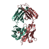 7u62C 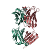 7u63C 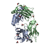 7u64C  2yk1S S: Starting model for refinement C: citing same article ( |
|---|---|
| Similar structure data |
-Links
 PDBj
PDBj

- Assembly
| Deposited unit | 
| ||||||||
|---|---|---|---|---|---|---|---|---|---|
| 1 |
| ||||||||
| Unit cell |
| ||||||||
| Components on special symmetry positions |
|
-Components
| #1: Antibody | Mass: 24107.164 Da / Num. of mol.: 1 / Source method: isolated from a natural source / Source: (natural) | ||||||
|---|---|---|---|---|---|---|---|
| #2: Antibody | Mass: 22833.430 Da / Num. of mol.: 1 / Source method: isolated from a natural source / Source: (natural) | ||||||
| #3: Chemical | ChemComp-NCT / (  Mass: 162.232 Da / Num. of mol.: 1 / Source method: obtained synthetically / Formula: C10H14N2 / Feature type: SUBJECT OF INVESTIGATION / Comment: alkaloid*YM Mass: 162.232 Da / Num. of mol.: 1 / Source method: obtained synthetically / Formula: C10H14N2 / Feature type: SUBJECT OF INVESTIGATION / Comment: alkaloid*YM | ||||||
| #4: Chemical |   Mass: 96.063 Da / Num. of mol.: 3 / Source method: obtained synthetically / Formula: SO4 Mass: 96.063 Da / Num. of mol.: 3 / Source method: obtained synthetically / Formula: SO4#5: Water | ChemComp-HOH / |  Mass: 18.015 Da / Num. of mol.: 89 / Source method: isolated from a natural source / Formula: H2O Mass: 18.015 Da / Num. of mol.: 89 / Source method: isolated from a natural source / Formula: H2OHas ligand of interest | Y | Has protein modification | Y | |
-Experimental details
-Experiment
| Experiment | Method: X-RAY DIFFRACTION / Number of used crystals: 1 |
|---|
- Sample preparation
| Crystal | Density Matthews: 2.45 Å3/Da / Density % sol: 49.8 % |
|---|---|
| Crystal grow | Temperature: 298 K / Method: vapor diffusion, sitting drop Details: MES pH 5.5, 60mM PEG 4000, 15% Ammonium Sulfate, 90mM |
-Data collection
| Diffraction | Mean temperature: 80 K / Serial crystal experiment: N | ||||||||||||||||||||||||||||||
|---|---|---|---|---|---|---|---|---|---|---|---|---|---|---|---|---|---|---|---|---|---|---|---|---|---|---|---|---|---|---|---|
| Diffraction source | Source: SYNCHROTRON / Site: ALS / Beamline: 5.0.2 / Wavelength: 1 Å | ||||||||||||||||||||||||||||||
| Detector | Type: DECTRIS PILATUS 6M / Detector: PIXEL / Date: Sep 6, 2018 | ||||||||||||||||||||||||||||||
| Radiation | Protocol: SINGLE WAVELENGTH / Monochromatic (M) / Laue (L): M / Scattering type: x-ray | ||||||||||||||||||||||||||||||
| Radiation wavelength | Wavelength: 1 Å / Relative weight: 1 | ||||||||||||||||||||||||||||||
| Reflection | Resolution: 2.05→59.74 Å / Num. obs: 27444 / % possible obs: 99 % / Redundancy: 2.8 % / Biso Wilson estimate: 34.46 Å2 / CC1/2: 0.999 / Rmerge(I) obs: 0.051 / Rpim(I) all: 0.036 / Rrim(I) all: 0.063 / Net I/σ(I): 23.5 / Num. measured all: 77729 / Scaling rejects: 4114 | ||||||||||||||||||||||||||||||
| Reflection shell | Diffraction-ID: 1
|
- Processing
| Software |
| ||||||||||||||||||||||||||||||||||||||||||||||||||||||||||||||||||||||||||||||||||||||||||||||||||
|---|---|---|---|---|---|---|---|---|---|---|---|---|---|---|---|---|---|---|---|---|---|---|---|---|---|---|---|---|---|---|---|---|---|---|---|---|---|---|---|---|---|---|---|---|---|---|---|---|---|---|---|---|---|---|---|---|---|---|---|---|---|---|---|---|---|---|---|---|---|---|---|---|---|---|---|---|---|---|---|---|---|---|---|---|---|---|---|---|---|---|---|---|---|---|---|---|---|---|---|
| Refinement | Method to determine structure: MOLECULAR REPLACEMENT Starting model: 2YK1 Resolution: 2.1→54.68 Å / SU ML: 0.29 / Cross valid method: THROUGHOUT / σ(F): 1.36 / Phase error: 30.27 / Stereochemistry target values: ML
| ||||||||||||||||||||||||||||||||||||||||||||||||||||||||||||||||||||||||||||||||||||||||||||||||||
| Solvent computation | Shrinkage radii: 0.9 Å / VDW probe radii: 1.11 Å / Solvent model: FLAT BULK SOLVENT MODEL | ||||||||||||||||||||||||||||||||||||||||||||||||||||||||||||||||||||||||||||||||||||||||||||||||||
| Displacement parameters | Biso max: 146.77 Å2 / Biso mean: 43.3249 Å2 / Biso min: 18.49 Å2 | ||||||||||||||||||||||||||||||||||||||||||||||||||||||||||||||||||||||||||||||||||||||||||||||||||
| Refinement step | Cycle: final / Resolution: 2.1→54.68 Å
| ||||||||||||||||||||||||||||||||||||||||||||||||||||||||||||||||||||||||||||||||||||||||||||||||||
| LS refinement shell | Refine-ID: X-RAY DIFFRACTION / Rfactor Rfree error: 0 / Total num. of bins used: 13
|
