 Movie
Movie Controller

[English] 日本語
 Yorodumi
- PDB-2yk1: Structure of human anti-nicotine Fab fragment in complex with nicotine -
+ Open data

- Basic information
| Entry | Database: PDB / ID: 2yk1 | ||||||
|---|---|---|---|---|---|---|---|
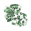
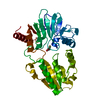
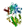
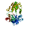
| Title | Structure of human anti-nicotine Fab fragment in complex with nicotine | ||||||
 Components Components |
| ||||||
 Keywords Keywords | IMMUNE SYSTEM / MONOCLONAL ANTIBODIES / ANTI-SMOKING VACCINE | ||||||
| Function / homology | Immunoglobulins / Immunoglobulin-like / Sandwich / Mainly Beta / (S)-3-(1-METHYLPYRROLIDIN-2-YL)PYRIDINE Function and homology information Function and homology information | ||||||
| Biological species |  HOMO SAPIENS (human) HOMO SAPIENS (human) | ||||||
| Method |  X-RAY DIFFRACTION / SYNCHROTRON / MOLECULAR REPLACEMENT / Resolution: 1.85 Å X-RAY DIFFRACTION / SYNCHROTRON / MOLECULAR REPLACEMENT / Resolution: 1.85 Å | ||||||
 Authors Authors | Tars, K. / Kotelovica, S. / Lipowsky, G. / Bauer, M. / Beerli, R. / Bachmann, M. / Maurer, P. | ||||||
 Citation Citation | Journal: J.Mol.Biol. / Year: 2012 Title: Different Binding Modes of Free and Carrier-Protein-Coupled Nicotine in a Human Monoclonal Antibody. Authors: Tars, K. / Kotelovica, S. / Lipowsky, G. / Bauer, M. / Beerli, R. / Bachmann, M. / Maurer, P. | ||||||
| History |
|
- Structure visualization
| Structure viewer | Molecule: MolmilJmol/JSmol |
|---|
- Downloads & links
-Download
| PDBx/mmCIF format | 2yk1.cif.gz | 157 KB | Display | PDBx/mmCIF format |
|---|---|---|---|---|
| PDB format | pdb2yk1.ent.gz | 123.3 KB | Display | PDB format |
| PDBx/mmJSON format | 2yk1.json.gz | Tree view | PDBx/mmJSON format | |
| Others |  Other downloads Other downloads |
-Validation report
| Arichive directory | https://data.pdbj.org/pub/pdb/validation_reports/yk/2yk1ftp://data.pdbj.org/pub/pdb/validation_reports/yk/2yk1 | HTTPS FTP |
|---|
-Related structure data
| Related structure data |  2yklC  2bosS S: Starting model for refinement C: citing same article ( |
|---|---|
| Similar structure data |


-Links
 PDBj
PDBj

- Assembly
| Deposited unit | 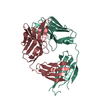
| |||||||||
|---|---|---|---|---|---|---|---|---|---|---|
| 1 |
| |||||||||
| Unit cell |
| |||||||||
| Components on special symmetry positions |
|
-Components
| #1: Antibody | Mass: 22297.004 Da / Num. of mol.: 1 Source method: isolated from a genetically manipulated source Source: (gene. exp.) HOMO SAPIENS (human) / Cell: B-CELL / Cell line (production host): HEK293T / Production host: HOMO SAPIENS (human) |
|---|---|
| #2: Antibody | Mass: 22976.256 Da / Num. of mol.: 1 Source method: isolated from a genetically manipulated source Source: (gene. exp.) HOMO SAPIENS (human) / Cell: B-CELL / Cell line (production host): HEK293T / Production host: HOMO SAPIENS (human) |
| #3: Chemical | ChemComp-NCT / (  Mass: 162.232 Da / Num. of mol.: 1 / Source method: obtained synthetically / Formula: C10H14N2 / Comment: alkaloid*YM Mass: 162.232 Da / Num. of mol.: 1 / Source method: obtained synthetically / Formula: C10H14N2 / Comment: alkaloid*YM |
| #4: Water | ChemComp-HOH /  Mass: 18.015 Da / Num. of mol.: 244 / Source method: isolated from a natural source / Formula: H2O Mass: 18.015 Da / Num. of mol.: 244 / Source method: isolated from a natural source / Formula: H2O |
| Has protein modification | Y |
-Experimental details
-Experiment
| Experiment | Method: X-RAY DIFFRACTION / Number of used crystals: 1 |
|---|
- Sample preparation
| Crystal | Density Matthews: 2.1 Å3/Da / Density % sol: 41 % / Description: NONE |
|---|---|
| Crystal grow | Method: vapor diffusion, sitting drop / pH: 6 Details: SITTING DROP, 5% PEG 3000, 0.1 M MES PH 6.0, 30% (V/V) PEG 200, 10 MG/ML PROTEIN, 20 MM NICOTINE. |
-Data collection
| Diffraction | Mean temperature: 100 K |
|---|---|
| Diffraction source | Source: SYNCHROTRON / Site: ESRF  / Beamline: ID14-1 / Wavelength: 0.933 / Beamline: ID14-1 / Wavelength: 0.933 |
| Detector | Type: ADSC QUANTUM 210 / Detector: CCD / Date: Oct 26, 2008 / Details: SAGITALLY FOCUSING GE(220) AND A MULTILAYER |
| Radiation | Monochromator: DIAMOND (111), GE(220) / Protocol: SINGLE WAVELENGTH / Monochromatic (M) / Laue (L): M / Scattering type: x-ray |
| Radiation wavelength | Wavelength: 0.933 Å / Relative weight: 1 |
| Reflection | Resolution: 1.85→30 Å / Num. obs: 34228 / % possible obs: 95 % / Observed criterion σ(I): 1.8 / Redundancy: 2.1 % / Biso Wilson estimate: 22 Å2 / Rmerge(I) obs: 0.04 / Net I/σ(I): 8.4 |
| Reflection shell | Resolution: 1.85→1.95 Å / Redundancy: 2.1 % / Rmerge(I) obs: 0.22 / Mean I/σ(I) obs: 1.8 / % possible all: 91.5 |
- Processing
| Software |
| ||||||||||||||||||||||||||||||||||||||||||||||||||||||||||||||||||||||||||||||||||||||||||||||||||||||||||||||||||||||||||||||||||||||||||||||||||||||||||||||||||||||||||||||||||||||
|---|---|---|---|---|---|---|---|---|---|---|---|---|---|---|---|---|---|---|---|---|---|---|---|---|---|---|---|---|---|---|---|---|---|---|---|---|---|---|---|---|---|---|---|---|---|---|---|---|---|---|---|---|---|---|---|---|---|---|---|---|---|---|---|---|---|---|---|---|---|---|---|---|---|---|---|---|---|---|---|---|---|---|---|---|---|---|---|---|---|---|---|---|---|---|---|---|---|---|---|---|---|---|---|---|---|---|---|---|---|---|---|---|---|---|---|---|---|---|---|---|---|---|---|---|---|---|---|---|---|---|---|---|---|---|---|---|---|---|---|---|---|---|---|---|---|---|---|---|---|---|---|---|---|---|---|---|---|---|---|---|---|---|---|---|---|---|---|---|---|---|---|---|---|---|---|---|---|---|---|---|---|---|---|
| Refinement | Method to determine structure: MOLECULAR REPLACEMENT Starting model: PDB ENTRY 2BOS Resolution: 1.85→27.35 Å / Cor.coef. Fo:Fc: 0.93 / Cor.coef. Fo:Fc free: 0.904 / SU B: 7.491 / SU ML: 0.107 / Cross valid method: THROUGHOUT / ESU R: 0.174 / ESU R Free: 0.159 / Stereochemistry target values: MAXIMUM LIKELIHOOD / Details: HYDROGENS HAVE BEEN ADDED IN THE RIDING POSITIONS.
| ||||||||||||||||||||||||||||||||||||||||||||||||||||||||||||||||||||||||||||||||||||||||||||||||||||||||||||||||||||||||||||||||||||||||||||||||||||||||||||||||||||||||||||||||||||||
| Solvent computation | Ion probe radii: 0.8 Å / Shrinkage radii: 0.8 Å / VDW probe radii: 1.4 Å / Solvent model: MASK | ||||||||||||||||||||||||||||||||||||||||||||||||||||||||||||||||||||||||||||||||||||||||||||||||||||||||||||||||||||||||||||||||||||||||||||||||||||||||||||||||||||||||||||||||||||||
| Displacement parameters | Biso mean: 28.267 Å2
| ||||||||||||||||||||||||||||||||||||||||||||||||||||||||||||||||||||||||||||||||||||||||||||||||||||||||||||||||||||||||||||||||||||||||||||||||||||||||||||||||||||||||||||||||||||||
| Refinement step | Cycle: LAST / Resolution: 1.85→27.35 Å
| ||||||||||||||||||||||||||||||||||||||||||||||||||||||||||||||||||||||||||||||||||||||||||||||||||||||||||||||||||||||||||||||||||||||||||||||||||||||||||||||||||||||||||||||||||||||
| Refine LS restraints |
|