 Movie
Movie Controller

[English] 日本語
 Yorodumi
- PDB-7u3b: Structure of S. venezuelae GlgX bound to c-di-GMP and acarbose (p... -
+ Open data

- Basic information
| Entry | Database: PDB / ID: 7u3b | ||||||
|---|---|---|---|---|---|---|---|
| Title | Structure of S. venezuelae GlgX bound to c-di-GMP and acarbose (pH 8.5) | ||||||
 Components Components | Glycogen debranching enzyme GlgX | ||||||
 Keywords Keywords | HYDROLASE / GlgX / glycogen / acarbose / c-di-GMP / Streptomyces | ||||||
| Function / homology |  Function and homology information Function and homology information: / glycogen catabolic process / hydrolase activity, hydrolyzing O-glycosyl compounds / nucleotide binding Similarity search - Function | ||||||
| Biological species |  Streptomyces venezuelae (bacteria) Streptomyces venezuelae (bacteria) | ||||||
| Method |  X-RAY DIFFRACTION / SYNCHROTRON / MOLECULAR REPLACEMENT / molecular replacement / Resolution: 3.6 Å X-RAY DIFFRACTION / SYNCHROTRON / MOLECULAR REPLACEMENT / molecular replacement / Resolution: 3.6 Å | ||||||
 Authors Authors | Schumacher, M.A. / Tschowri, N. | ||||||
| Funding support |  United States, 1items United States, 1items
| ||||||
 Citation Citation | Journal: Nat Commun / Year: 2022 Title: Allosteric regulation of glycogen breakdown by the second messenger cyclic di-GMP. Authors: Schumacher, M.A. / Wormann, M.E. / Henderson, M. / Salinas, R. / Latoscha, A. / Al-Bassam, M.M. / Singh, K.S. / Barclay, E. / Gunka, K. / Tschowri, N. | ||||||
| History |
|
- Structure visualization
| Structure viewer | Molecule: MolmilJmol/JSmol |
|---|
- Downloads & links
-Download
| PDBx/mmCIF format | 7u3b.cif.gz | 1.1 MB | Display | PDBx/mmCIF format |
|---|---|---|---|---|
| PDB format | pdb7u3b.ent.gz | 895.4 KB | Display | PDB format |
| PDBx/mmJSON format | 7u3b.json.gz | Tree view | PDBx/mmJSON format | |
| Others |  Other downloads Other downloads |
-Validation report
| Summary document | 7u3b_validation.pdf.gz | 5.2 MB | Display | wwPDB validaton report |
|---|---|---|---|---|
| Full document | 7u3b_full_validation.pdf.gz | 5.3 MB | Display | |
| Data in XML | 7u3b_validation.xml.gz | 220.3 KB | Display | |
| Data in CIF | 7u3b_validation.cif.gz | 279.5 KB | Display | |
| Arichive directory | https://data.pdbj.org/pub/pdb/validation_reports/u3/7u3bftp://data.pdbj.org/pub/pdb/validation_reports/u3/7u3b | HTTPS FTP |
-Related structure data
| Related structure data |  7u39C  7u3aSC  7u3dC S: Starting model for refinement C: citing same article ( |
|---|---|
| Similar structure data |
-Links
 PDBj
PDBj


- Assembly
| Deposited unit | 
| ||||||||
|---|---|---|---|---|---|---|---|---|---|
| 1 | 
| ||||||||
| 2 | 
| ||||||||
| 3 | 
| ||||||||
| 4 | 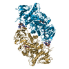
| ||||||||
| Unit cell |
|
-Components
| #1: Protein | Mass: 80167.234 Da / Num. of mol.: 8 Source method: isolated from a genetically manipulated source Source: (gene. exp.) Streptomyces venezuelae (bacteria) / Gene: glgX, DEJ46_08920 / Production host: #2: Chemical | ChemComp-A16 /   Mass: 485.480 Da / Num. of mol.: 8 / Source method: obtained synthetically / Formula: C19H35NO13 / Feature type: SUBJECT OF INVESTIGATION Mass: 485.480 Da / Num. of mol.: 8 / Source method: obtained synthetically / Formula: C19H35NO13 / Feature type: SUBJECT OF INVESTIGATION#3: Chemical | ChemComp-C2E /   Mass: 690.411 Da / Num. of mol.: 8 / Source method: obtained synthetically / Formula: C20H24N10O14P2 Mass: 690.411 Da / Num. of mol.: 8 / Source method: obtained synthetically / Formula: C20H24N10O14P2#4: Chemical | ChemComp-PO4 /   Mass: 94.971 Da / Num. of mol.: 4 / Source method: obtained synthetically / Formula: PO4 Mass: 94.971 Da / Num. of mol.: 4 / Source method: obtained synthetically / Formula: PO4#5: Water | ChemComp-HOH / |  Mass: 18.015 Da / Num. of mol.: 20 / Source method: isolated from a natural source / Formula: H2O Mass: 18.015 Da / Num. of mol.: 20 / Source method: isolated from a natural source / Formula: H2OHas ligand of interest | Y | Has protein modification | N | |
|---|
-Experimental details
-Experiment
| Experiment | Method: X-RAY DIFFRACTION / Number of used crystals: 1 |
|---|
- Sample preparation
| Crystal | Density Matthews: 2.91 Å3/Da / Density % sol: 57.67 % |
|---|---|
| Crystal grow | Temperature: 298 K / Method: vapor diffusion, hanging drop Details: 50% MPD, 0.1 M Tris pH 8.5 and 200 mM ammonium phosphate monobasic |
-Data collection
| Diffraction | Mean temperature: 100 K / Serial crystal experiment: N |
|---|---|
| Diffraction source | Source: SYNCHROTRON / Site: ALS / Beamline: 5.0.1 / Wavelength: 1 Å |
| Detector | Type: DECTRIS PILATUS3 2M / Detector: PIXEL / Date: Jan 12, 2021 |
| Radiation | Protocol: SINGLE WAVELENGTH / Monochromatic (M) / Laue (L): M / Scattering type: x-ray |
| Radiation wavelength | Wavelength: 1 Å / Relative weight: 1 |
| Reflection | Resolution: 3.6→48.5 Å / Num. obs: 83324 / % possible obs: 98.2 % / Redundancy: 1.7 % / CC1/2: 0.923 / Rpim(I) all: 0.17 / Rsym value: 0.199 / Net I/σ(I): 5.5 |
| Reflection shell | Resolution: 3.6→3.78 Å / Mean I/σ(I) obs: 1.3 / Num. unique obs: 5752 / CC1/2: 0.667 / Rpim(I) all: 0.517 / Rsym value: 0.78 |
-Phasing
| Phasing | Method: molecular replacement |
|---|
- Processing
| Software |
| |||||||||||||||||||||||||||||||||||||||||||||||||||||||||||||||||||||||||||||||||||||||||||||||||||||||||
|---|---|---|---|---|---|---|---|---|---|---|---|---|---|---|---|---|---|---|---|---|---|---|---|---|---|---|---|---|---|---|---|---|---|---|---|---|---|---|---|---|---|---|---|---|---|---|---|---|---|---|---|---|---|---|---|---|---|---|---|---|---|---|---|---|---|---|---|---|---|---|---|---|---|---|---|---|---|---|---|---|---|---|---|---|---|---|---|---|---|---|---|---|---|---|---|---|---|---|---|---|---|---|---|---|---|---|
| Refinement | Method to determine structure: MOLECULAR REPLACEMENT Starting model: 7U3A Resolution: 3.6→48.5 Å / SU ML: 0.5 / Cross valid method: THROUGHOUT / σ(F): 1.35 / Phase error: 30.67 / Stereochemistry target values: ML
| |||||||||||||||||||||||||||||||||||||||||||||||||||||||||||||||||||||||||||||||||||||||||||||||||||||||||
| Solvent computation | Shrinkage radii: 0.9 Å / VDW probe radii: 1.11 Å / Solvent model: FLAT BULK SOLVENT MODEL | |||||||||||||||||||||||||||||||||||||||||||||||||||||||||||||||||||||||||||||||||||||||||||||||||||||||||
| Displacement parameters | Biso max: 216.7 Å2 / Biso mean: 46.8686 Å2 / Biso min: 0.12 Å2 | |||||||||||||||||||||||||||||||||||||||||||||||||||||||||||||||||||||||||||||||||||||||||||||||||||||||||
| Refinement step | Cycle: final / Resolution: 3.6→48.5 Å
| |||||||||||||||||||||||||||||||||||||||||||||||||||||||||||||||||||||||||||||||||||||||||||||||||||||||||
| LS refinement shell | Refine-ID: X-RAY DIFFRACTION / Rfactor Rfree error: 0 / Total num. of bins used: 14
|
