 Movie
Movie Controller


+ Open data

- Basic information
| Entry | Database: PDB / ID: 7u3d | ||||||
|---|---|---|---|---|---|---|---|
| Title | Structure of S. venezuelae GlgX-c-di-GMP-acarbose complex (4.6) | ||||||
 Components Components | Glycogen debranching enzyme GlgX | ||||||
 Keywords Keywords | HYDROLASE / GlgX / glycogen / c-di-GMP / acarbose / Streptomyces development | ||||||
| Function / homology |  Function and homology information Function and homology informationamylo-alpha-1,6-glucosidase activity / glycogen catabolic process / nucleotide binding Similarity search - Function | ||||||
| Biological species |  Streptomyces venezuelae (bacteria) Streptomyces venezuelae (bacteria) | ||||||
| Method |  X-RAY DIFFRACTION / SYNCHROTRON / MOLECULAR REPLACEMENT / molecular replacement / Resolution: 2.4 Å X-RAY DIFFRACTION / SYNCHROTRON / MOLECULAR REPLACEMENT / molecular replacement / Resolution: 2.4 Å | ||||||
 Authors Authors | Schumacher, M.A. | ||||||
| Funding support |  United States, 1items United States, 1items
| ||||||
 Citation Citation | Journal: Nat Commun / Year: 2022 Title: Allosteric regulation of glycogen breakdown by the second messenger cyclic di-GMP. Authors: Schumacher, M.A. / Wormann, M.E. / Henderson, M. / Salinas, R. / Latoscha, A. / Al-Bassam, M.M. / Singh, K.S. / Barclay, E. / Gunka, K. / Tschowri, N. | ||||||
| History |
|
- Structure visualization
| Structure viewer | Molecule: MolmilJmol/JSmol |
|---|
- Downloads & links
-Download
| PDBx/mmCIF format | 7u3d.cif.gz | 1.1 MB | Display | PDBx/mmCIF format |
|---|---|---|---|---|
| PDB format | pdb7u3d.ent.gz | 926.8 KB | Display | PDB format |
| PDBx/mmJSON format | 7u3d.json.gz | Tree view | PDBx/mmJSON format | |
| Others |  Other downloads Other downloads |
-Validation report
| Arichive directory | https://data.pdbj.org/pub/pdb/validation_reports/u3/7u3dftp://data.pdbj.org/pub/pdb/validation_reports/u3/7u3d | HTTPS FTP |
|---|
-Related structure data
| Related structure data | 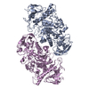 7u39C  7u3aC  7u3bSC S: Starting model for refinement C: citing same article ( |
|---|---|
| Similar structure data |
-Links
 PDBj
PDBj


- Assembly
| Deposited unit | 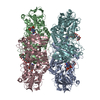
| ||||||||
|---|---|---|---|---|---|---|---|---|---|
| 1 | 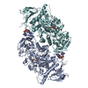
| ||||||||
| 2 | 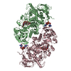
| ||||||||
| Unit cell |
| ||||||||
| Components on special symmetry positions |
|
-Components
| #1: Protein | Mass: 80167.234 Da / Num. of mol.: 4 Source method: isolated from a genetically manipulated source Source: (gene. exp.) Streptomyces venezuelae (bacteria) / Gene: glgX, DEJ46_08920 / Production host: #2: Polysaccharide | 4,6-dideoxy-4-{[(1S,4R,5S,6S)-4,5,6-trihydroxy-3-(hydroxymethyl)cyclohex-2-en-1-yl]amino}-alpha-D- ...4,6-dideoxy-4-{[(1S,4R,5S,6S)-4,5,6-trihydroxy-3-(hydroxymethyl)cyclohex-2-en-1-yl]amino}-alpha-D-glucopyranose-(1-4)-alpha-D-glucopyranose-(1-4)-alpha-D-glucopyranose   Type: oligosaccharide, Oligosaccharide / Class: Inhibitor / Mass: 645.606 Da / Num. of mol.: 4 Type: oligosaccharide, Oligosaccharide / Class: Inhibitor / Mass: 645.606 Da / Num. of mol.: 4Source method: isolated from a genetically manipulated source Details: oligosaccharide / References: alpha-acarbose #3: Chemical | ChemComp-C2E /   Mass: 690.411 Da / Num. of mol.: 4 / Source method: obtained synthetically / Formula: C20H24N10O14P2 / Feature type: SUBJECT OF INVESTIGATION Mass: 690.411 Da / Num. of mol.: 4 / Source method: obtained synthetically / Formula: C20H24N10O14P2 / Feature type: SUBJECT OF INVESTIGATION#4: Water | ChemComp-HOH / |  Mass: 18.015 Da / Num. of mol.: 1016 / Source method: isolated from a natural source / Formula: H2O Mass: 18.015 Da / Num. of mol.: 1016 / Source method: isolated from a natural source / Formula: H2OHas ligand of interest | Y | |
|---|
-Experimental details
-Experiment
| Experiment | Method: X-RAY DIFFRACTION / Number of used crystals: 1 |
|---|
- Sample preparation
| Crystal | Density Matthews: 2.47 Å3/Da / Density % sol: 50.15 % |
|---|---|
| Crystal grow | Temperature: 298 K / Method: vapor diffusion, hanging drop Details: 30% PEG 300, 100 mM NaCl and 0.1 sodium acetate pH 4.6 |
-Data collection
| Diffraction | Mean temperature: 100 K / Serial crystal experiment: N |
|---|---|
| Diffraction source | Source: SYNCHROTRON / Site: ALS / Beamline: 5.0.2 / Wavelength: 1 Å |
| Detector | Type: DECTRIS PILATUS3 6M / Detector: PIXEL / Date: Feb 3, 2020 |
| Radiation | Protocol: SINGLE WAVELENGTH / Monochromatic (M) / Laue (L): M / Scattering type: x-ray |
| Radiation wavelength | Wavelength: 1 Å / Relative weight: 1 |
| Reflection | Resolution: 2.4→49.1 Å / Num. obs: 124127 / % possible obs: 99.9 % / Redundancy: 6.5 % / CC1/2: 0.995 / Rpim(I) all: 0.07 / Rsym value: 0.155 / Net I/σ(I): 11 |
| Reflection shell | Resolution: 2.4→2.53 Å / Mean I/σ(I) obs: 1.9 / Num. unique obs: 8764 / CC1/2: 0.647 / Rpim(I) all: 0.0473 / Rsym value: 1.045 |
-Phasing
| Phasing | Method: molecular replacement |
|---|
- Processing
| Software |
| |||||||||||||||||||||||||||||||||||||||||||||||||||||||||||||||||||||||||||||||||||||||||||||||||||||||||
|---|---|---|---|---|---|---|---|---|---|---|---|---|---|---|---|---|---|---|---|---|---|---|---|---|---|---|---|---|---|---|---|---|---|---|---|---|---|---|---|---|---|---|---|---|---|---|---|---|---|---|---|---|---|---|---|---|---|---|---|---|---|---|---|---|---|---|---|---|---|---|---|---|---|---|---|---|---|---|---|---|---|---|---|---|---|---|---|---|---|---|---|---|---|---|---|---|---|---|---|---|---|---|---|---|---|---|
| Refinement | Method to determine structure: MOLECULAR REPLACEMENT Starting model: 7U3B Resolution: 2.4→47.41 Å / SU ML: 0.32 / Cross valid method: THROUGHOUT / σ(F): 1.33 / Phase error: 24.51 / Stereochemistry target values: ML
| |||||||||||||||||||||||||||||||||||||||||||||||||||||||||||||||||||||||||||||||||||||||||||||||||||||||||
| Solvent computation | Shrinkage radii: 0.9 Å / VDW probe radii: 1.11 Å / Solvent model: FLAT BULK SOLVENT MODEL | |||||||||||||||||||||||||||||||||||||||||||||||||||||||||||||||||||||||||||||||||||||||||||||||||||||||||
| Displacement parameters | Biso max: 134.69 Å2 / Biso mean: 39.7524 Å2 / Biso min: 19.19 Å2 | |||||||||||||||||||||||||||||||||||||||||||||||||||||||||||||||||||||||||||||||||||||||||||||||||||||||||
| Refinement step | Cycle: final / Resolution: 2.4→47.41 Å
| |||||||||||||||||||||||||||||||||||||||||||||||||||||||||||||||||||||||||||||||||||||||||||||||||||||||||
| LS refinement shell | Refine-ID: X-RAY DIFFRACTION / Rfactor Rfree error: 0 / Total num. of bins used: 14
| |||||||||||||||||||||||||||||||||||||||||||||||||||||||||||||||||||||||||||||||||||||||||||||||||||||||||
| Refinement TLS params. | Method: refined / Origin x: 14.4425 Å / Origin y: -37.319 Å / Origin z: 28.1626 Å
| |||||||||||||||||||||||||||||||||||||||||||||||||||||||||||||||||||||||||||||||||||||||||||||||||||||||||
| Refinement TLS group |
|
