 Movie
Movie Controller

[English] 日本語
 Yorodumi
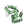
- PDB-7u1n: Crystal structure of the Anopheles darlingi AD-118 long form D7 s... -
+ Open data

- Basic information
| Entry | Database: PDB / ID: 7u1n | ||||||
|---|---|---|---|---|---|---|---|
| Title | Crystal structure of the Anopheles darlingi AD-118 long form D7 salivary protein | ||||||
 Components Components | Long form D7 salivary protein | ||||||
 Keywords Keywords | BLOOD CLOTTING / odorant binding protein family / blood feeding | ||||||
| Function / homology | Pheromone/general odorant binding protein superfamily / odorant binding / sensory perception of smell / toxin activity / : / 2-ETHOXYETHANOL / DI(HYDROXYETHYL)ETHER / Long form salivary protein D7L2 Function and homology information Function and homology information | ||||||
| Biological species |  | ||||||
| Method |  X-RAY DIFFRACTION / MIRAS / Resolution: 2.4 Å X-RAY DIFFRACTION / MIRAS / Resolution: 2.4 Å | ||||||
 Authors Authors | Alvarenga, P.H. / Gittis, A.G. / Garboczi, D.N. / Andersen, J.F. | ||||||
| Funding support |  United States, 1items United States, 1items
| ||||||
 Citation Citation | Journal: Insect Biochem.Mol.Biol. / Year: 2022 Title: Functional aspects of evolution in a cluster of salivary protein genes from mosquitoes. Authors: Alvarenga, P.H. / Dias, D.R. / Xu, X. / Francischetti, I.M.B. / Gittis, A.G. / Arp, G. / Garboczi, D.N. / Ribeiro, J.M.C. / Andersen, J.F. | ||||||
| History |
|
- Structure visualization
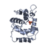
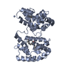
| Structure viewer | Molecule: MolmilJmol/JSmol |
|---|
- Downloads & links
-Download
| PDBx/mmCIF format | 7u1n.cif.gz | 143.3 KB | Display | PDBx/mmCIF format |
|---|---|---|---|---|
| PDB format | pdb7u1n.ent.gz | 100.8 KB | Display | PDB format |
| PDBx/mmJSON format | 7u1n.json.gz | Tree view | PDBx/mmJSON format | |
| Others |  Other downloads Other downloads |
-Validation report
| Arichive directory | https://data.pdbj.org/pub/pdb/validation_reports/u1/7u1nftp://data.pdbj.org/pub/pdb/validation_reports/u1/7u1n | HTTPS FTP |
|---|
-Related structure data
-Links
 PDBj
PDBj- Assembly
| Deposited unit | 
| ||||||||||||
|---|---|---|---|---|---|---|---|---|---|---|---|---|---|
| 1 | 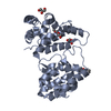
| ||||||||||||
| 2 | 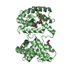
| ||||||||||||
| Unit cell |
|
-Components
-Protein , 1 types, 2 molecules AB
| #1: Protein | Mass: 34892.090 Da / Num. of mol.: 2 Source method: isolated from a genetically manipulated source Source: (gene. exp.) Production host:  |
|---|
-Non-polymers , 7 types, 137 molecules 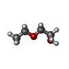


| #2: Chemical |  Mass: 90.121 Da / Num. of mol.: 2 / Source method: obtained synthetically / Formula: C4H10O2 Mass: 90.121 Da / Num. of mol.: 2 / Source method: obtained synthetically / Formula: C4H10O2#3: Chemical |  Mass: 96.063 Da / Num. of mol.: 2 / Source method: obtained synthetically / Formula: SO4 Mass: 96.063 Da / Num. of mol.: 2 / Source method: obtained synthetically / Formula: SO4#4: Chemical |  Mass: 120.147 Da / Num. of mol.: 2 / Source method: obtained synthetically / Formula: C5H12O3 / Comment: inhibitor, precipitant*YM Mass: 120.147 Da / Num. of mol.: 2 / Source method: obtained synthetically / Formula: C5H12O3 / Comment: inhibitor, precipitant*YM#5: Chemical |  Mass: 92.094 Da / Num. of mol.: 3 / Source method: obtained synthetically / Formula: C3H8O3 Mass: 92.094 Da / Num. of mol.: 3 / Source method: obtained synthetically / Formula: C3H8O3#6: Chemical | ChemComp-PEG / |  Mass: 106.120 Da / Num. of mol.: 1 / Source method: obtained synthetically / Formula: C4H10O3 Mass: 106.120 Da / Num. of mol.: 1 / Source method: obtained synthetically / Formula: C4H10O3#7: Chemical | ChemComp-EDO / |  Mass: 62.068 Da / Num. of mol.: 1 / Source method: obtained synthetically / Formula: C2H6O2 Mass: 62.068 Da / Num. of mol.: 1 / Source method: obtained synthetically / Formula: C2H6O2#8: Water | ChemComp-HOH / | Mass: 18.015 Da / Num. of mol.: 126 / Source method: isolated from a natural source / Formula: H2O |
|---|
-Details
| Has ligand of interest | N |
|---|---|
| Has protein modification | Y |
-Experimental details
-Experiment
| Experiment | Method: X-RAY DIFFRACTION / Number of used crystals: 1 |
|---|
- Sample preparation
| Crystal | Density Matthews: 2.43 Å3/Da / Density % sol: 49.32 % |
|---|---|
| Crystal grow | Temperature: 298 K / Method: vapor diffusion / pH: 7.5 Details: 30%P EG 5000 MME, 0.2 M ammonium sulfate, 0.1M HEPES |
-Data collection
| Diffraction | Mean temperature: 95 K / Serial crystal experiment: N |
|---|---|
| Diffraction source | Source: ROTATING ANODE / Type: RIGAKU / Wavelength: 1.54178 Å |
| Detector | Type: RIGAKU SATURN A200 / Detector: CCD / Date: Aug 12, 2020 |
| Radiation | Protocol: SINGLE WAVELENGTH / Monochromatic (M) / Laue (L): M / Scattering type: x-ray |
| Radiation wavelength | Wavelength: 1.54178 Å / Relative weight: 1 |
| Reflection | Resolution: 2.4→29.39 Å / Num. obs: 24809 / % possible obs: 94.1 % / Redundancy: 4.78 % / Biso Wilson estimate: 27.19 Å2 / Rmerge(I) obs: 0.065 / Net I/σ(I): 15.77 |
| Reflection shell | Resolution: 2.4→2.46 Å / Rmerge(I) obs: 0.284 / Num. unique obs: 1239 / % possible all: 63.4 |
- Processing
| Software |
| |||||||||||||||||||||||||||||||||||||||||||||||||||||||||||||||||||||||||||||||||||||||||||||||||||||||||
|---|---|---|---|---|---|---|---|---|---|---|---|---|---|---|---|---|---|---|---|---|---|---|---|---|---|---|---|---|---|---|---|---|---|---|---|---|---|---|---|---|---|---|---|---|---|---|---|---|---|---|---|---|---|---|---|---|---|---|---|---|---|---|---|---|---|---|---|---|---|---|---|---|---|---|---|---|---|---|---|---|---|---|---|---|---|---|---|---|---|---|---|---|---|---|---|---|---|---|---|---|---|---|---|---|---|---|
| Refinement | Method to determine structure: MIRAS / Resolution: 2.4→29.39 Å / SU ML: 0.3149 / Cross valid method: FREE R-VALUE / σ(F): 1.99 / Phase error: 33.713 Stereochemistry target values: GeoStd + Monomer Library + CDL v1.2
| |||||||||||||||||||||||||||||||||||||||||||||||||||||||||||||||||||||||||||||||||||||||||||||||||||||||||
| Solvent computation | Shrinkage radii: 0.9 Å / VDW probe radii: 1.11 Å / Solvent model: FLAT BULK SOLVENT MODEL | |||||||||||||||||||||||||||||||||||||||||||||||||||||||||||||||||||||||||||||||||||||||||||||||||||||||||
| Displacement parameters | Biso mean: 30.84 Å2 | |||||||||||||||||||||||||||||||||||||||||||||||||||||||||||||||||||||||||||||||||||||||||||||||||||||||||
| Refinement step | Cycle: LAST / Resolution: 2.4→29.39 Å
| |||||||||||||||||||||||||||||||||||||||||||||||||||||||||||||||||||||||||||||||||||||||||||||||||||||||||
| Refine LS restraints |
| |||||||||||||||||||||||||||||||||||||||||||||||||||||||||||||||||||||||||||||||||||||||||||||||||||||||||
| LS refinement shell |
|
