 Movie
Movie Controller


+ Open data

- Basic information
| Entry | Database: PDB / ID: 7tug | ||||||
|---|---|---|---|---|---|---|---|
| Title | Crystal structure of Tapasin in complex with PaSta2-Fab | ||||||
 Components Components |
| ||||||
 Keywords Keywords | IMMUNE SYSTEM / PaSta / Fab / Antibody / IgG / MHC-I / HLA / peptide loading complex / PLC / antigen presentation / immune response | ||||||
| Function / homology |  Function and homology information Function and homology informationMHC class Ib protein complex assembly / peptide antigen stabilization / Tapasin-ERp57 complex / MHC class I protein complex binding / antigen processing and presentation of exogenous peptide antigen via MHC class I, TAP-dependent / TAP2 binding / TAP1 binding / regulation of protein complex stability / retrograde vesicle-mediated transport, Golgi to endoplasmic reticulum / TAP complex binding ...MHC class Ib protein complex assembly / peptide antigen stabilization / Tapasin-ERp57 complex / MHC class I protein complex binding / antigen processing and presentation of exogenous peptide antigen via MHC class I, TAP-dependent / TAP2 binding / TAP1 binding / regulation of protein complex stability / retrograde vesicle-mediated transport, Golgi to endoplasmic reticulum / TAP complex binding / MHC class I protein binding / endoplasmic reticulum-Golgi intermediate compartment membrane / protein folding chaperone / Antigen Presentation: Folding, assembly and peptide loading of class I MHC / lumenal side of endoplasmic reticulum membrane / peptide antigen assembly with MHC class I protein complex / MHC class I peptide loading complex / antigen processing and presentation of endogenous peptide antigen via MHC class I / peptide antigen binding / phagocytic vesicle membrane / : / ER-Phagosome pathway / regulation of gene expression / protein-containing complex assembly / molecular adaptor activity / Golgi membrane / endoplasmic reticulum membrane / endoplasmic reticulum Similarity search - Function | ||||||
| Biological species |  Homo sapiens (human) Homo sapiens (human) | ||||||
| Method |  X-RAY DIFFRACTION / SYNCHROTRON / MOLECULAR REPLACEMENT / Resolution: 3.9 Å X-RAY DIFFRACTION / SYNCHROTRON / MOLECULAR REPLACEMENT / Resolution: 3.9 Å | ||||||
 Authors Authors | Jiang, J. / Natarajan, K. / Taylor, D.K. / Boyd, L.F. / Margulies, D.H. | ||||||
| Funding support |  United States, 1items United States, 1items
| ||||||
 Citation Citation | Journal: Nat Commun / Year: 2022 Title: Structural mechanism of tapasin-mediated MHC-I peptide loading in antigen presentation. Authors: Jiang, J. / Taylor, D.K. / Kim, E.J. / Boyd, L.F. / Ahmad, J. / Mage, M.G. / Truong, H.V. / Woodward, C.H. / Sgourakis, N.G. / Cresswell, P. / Margulies, D.H. / Natarajan, K. | ||||||
| History |
|
- Structure visualization
| Structure viewer | Molecule: MolmilJmol/JSmol |
|---|
- Downloads & links
-Download
| PDBx/mmCIF format | 7tug.cif.gz | 186.7 KB | Display | PDBx/mmCIF format |
|---|---|---|---|---|
| PDB format | pdb7tug.ent.gz | 115.9 KB | Display | PDB format |
| PDBx/mmJSON format | 7tug.json.gz | Tree view | PDBx/mmJSON format | |
| Others |  Other downloads Other downloads |
-Validation report
| Arichive directory | https://data.pdbj.org/pub/pdb/validation_reports/tu/7tugftp://data.pdbj.org/pub/pdb/validation_reports/tu/7tug | HTTPS FTP |
|---|
-Related structure data
| Related structure data |  7tucC  7tudC  7tueC  7tufC 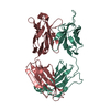 7tuhC  3f8uS C: citing same article ( S: Starting model for refinement |
|---|---|
| Similar structure data |
-Links
 PDBj
PDBj

- Assembly
| Deposited unit | 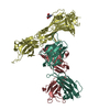
| ||||||||||||
|---|---|---|---|---|---|---|---|---|---|---|---|---|---|
| 1 | 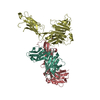
| ||||||||||||
| Unit cell |
|
-Components
| #1: Antibody | Mass: 44739.641 Da / Num. of mol.: 1 Source method: isolated from a genetically manipulated source Source: (gene. exp.) Homo sapiens (human) / Gene: TAPBP, NGS17, TAPA / Plasmid: pET21b / Cell line (production host): S2 / Production host:  |
|---|---|
| #2: Antibody | Mass: 24792.748 Da / Num. of mol.: 1 / Fragment: Variable and Constant CH1 Source method: isolated from a genetically manipulated source Source: (gene. exp.) Homo sapiens (human) |
| #3: Antibody | Mass: 24278.922 Da / Num. of mol.: 1 Source method: isolated from a genetically manipulated source Source: (gene. exp.) Homo sapiens (human) |
| #4: Sugar | ChemComp-NAG /   Type: D-saccharide, beta linking / Mass: 221.208 Da / Num. of mol.: 1 / Source method: obtained synthetically / Formula: C8H15NO6 Type: D-saccharide, beta linking / Mass: 221.208 Da / Num. of mol.: 1 / Source method: obtained synthetically / Formula: C8H15NO6 |
| Has ligand of interest | N |
| Has protein modification | Y |
-Experimental details
-Experiment
| Experiment | Method: X-RAY DIFFRACTION / Number of used crystals: 1 |
|---|
- Sample preparation
| Crystal | Density Matthews: 2.47 Å3/Da / Density % sol: 50.22 % |
|---|---|
| Crystal grow | Temperature: 291 K / Method: vapor diffusion, hanging drop / pH: 5.5 / Details: 17% PEG 10000, 0.1M Bis-Tris, 0.1M Am Acetate / PH range: 5.5-8.5 |
-Data collection
| Diffraction | Mean temperature: 273 K / Ambient temp details: LN blow / Serial crystal experiment: N |
|---|---|
| Diffraction source | Source: SYNCHROTRON / Site: APS / Beamline: 22-ID / Wavelength: 1 Å |
| Detector | Type: DECTRIS EIGER X 16M / Detector: PIXEL / Date: Jun 20, 2021 |
| Radiation | Protocol: SINGLE WAVELENGTH / Monochromatic (M) / Laue (L): M / Scattering type: x-ray |
| Radiation wavelength | Wavelength: 1 Å / Relative weight: 1 |
| Reflection | Resolution: 3.9→54.33 Å / Num. obs: 8241 / % possible obs: 95.4 % / Redundancy: 3.5 % / Biso Wilson estimate: 67.65 Å2 / CC1/2: 0.998 / Rmerge(I) obs: 0.107 / Rpim(I) all: 0.065 / Rrim(I) all: 0.127 / Net I/σ(I): 7.5 |
| Reflection shell | Resolution: 3.9→4.04 Å / Mean I/σ(I) obs: 0.85 / Num. unique obs: 692 / CC1/2: 0.477 / Rpim(I) all: 0.851 / % possible all: 80.75 |
- Processing
| Software |
| ||||||||||||||||||||||||||||
|---|---|---|---|---|---|---|---|---|---|---|---|---|---|---|---|---|---|---|---|---|---|---|---|---|---|---|---|---|---|
| Refinement | Method to determine structure: MOLECULAR REPLACEMENT Starting model: 3F8U Resolution: 3.9→54.33 Å / SU ML: 0.6412 / Cross valid method: FREE R-VALUE / σ(F): 2 / Phase error: 34.8533 Stereochemistry target values: GeoStd + Monomer Library + CDL v1.2
| ||||||||||||||||||||||||||||
| Solvent computation | Shrinkage radii: 0.9 Å / VDW probe radii: 1.11 Å / Solvent model: FLAT BULK SOLVENT MODEL | ||||||||||||||||||||||||||||
| Displacement parameters | Biso mean: 78.23 Å2 | ||||||||||||||||||||||||||||
| Refinement step | Cycle: LAST / Resolution: 3.9→54.33 Å
| ||||||||||||||||||||||||||||
| Refine LS restraints |
| ||||||||||||||||||||||||||||
| LS refinement shell |
|
