[English] 日本語
 Yorodumi
- PDB-7tr3: CaKip3[2-482] - AMP-PNP in complex with a dolastatin-10-stabilize... -
+ Open data
- Basic information
| Entry | Database: PDB / ID: 7tr3 | ||||||||||||
|---|---|---|---|---|---|---|---|---|---|---|---|---|---|
| Title | CaKip3[2-482] - AMP-PNP in complex with a dolastatin-10-stabilized tubulin ring | ||||||||||||
 Components Components |
| ||||||||||||
 Keywords Keywords | MOTOR PROTEIN / Kip3 / kinesin / tubulin / dolastatin-10 | ||||||||||||
| Function / homology |  Function and homology information Function and homology informationplus-end specific microtubule depolymerization / tubulin-dependent ATPase activity / regulation of mitotic spindle elongation / meiotic sister chromatid segregation / mitotic spindle astral microtubule / mitotic spindle midzone / nuclear microtubule / nuclear migration along microtubule / microtubule plus-end / microtubule nucleation ...plus-end specific microtubule depolymerization / tubulin-dependent ATPase activity / regulation of mitotic spindle elongation / meiotic sister chromatid segregation / mitotic spindle astral microtubule / mitotic spindle midzone / nuclear microtubule / nuclear migration along microtubule / microtubule plus-end / microtubule nucleation / plus-end-directed microtubule motor activity / mitotic spindle disassembly / Microtubule-dependent trafficking of connexons from Golgi to the plasma membrane / Resolution of Sister Chromatid Cohesion / Hedgehog 'off' state / Cilium Assembly / Intraflagellar transport / COPI-dependent Golgi-to-ER retrograde traffic / Mitotic Prometaphase / Carboxyterminal post-translational modifications of tubulin / RHOH GTPase cycle / EML4 and NUDC in mitotic spindle formation / Sealing of the nuclear envelope (NE) by ESCRT-III / Kinesins / PKR-mediated signaling / Separation of Sister Chromatids / The role of GTSE1 in G2/M progression after G2 checkpoint / Aggrephagy / kinesin complex / microtubule depolymerization / RHO GTPases activate IQGAPs / RHO GTPases Activate Formins / HSP90 chaperone cycle for steroid hormone receptors (SHR) in the presence of ligand / MHC class II antigen presentation / Recruitment of NuMA to mitotic centrosomes / COPI-mediated anterograde transport / microtubule-based movement / negative regulation of microtubule polymerization / establishment of mitotic spindle orientation / mitotic sister chromatid segregation / mitotic spindle assembly / microtubule-based process / mitotic spindle organization / structural constituent of cytoskeleton / microtubule cytoskeleton organization / mitotic cell cycle / microtubule cytoskeleton / microtubule binding / Hydrolases; Acting on acid anhydrides; Acting on GTP to facilitate cellular and subcellular movement / microtubule / GTPase activity / GTP binding / ATP hydrolysis activity / ATP binding / metal ion binding / nucleus / cytoplasm Similarity search - Function | ||||||||||||
| Biological species |  Candida albicans (yeast) Candida albicans (yeast) | ||||||||||||
| Method | ELECTRON MICROSCOPY / single particle reconstruction / cryo EM / Resolution: 3.9 Å | ||||||||||||
 Authors Authors | Benoit, M.P.M.H. / Asenjo, A.B. / Hunter, B. / Allingham, J.S. / Sosa, H. | ||||||||||||
| Funding support |  United States, United States,  Canada, 3items Canada, 3items
| ||||||||||||
 Citation Citation |  Journal: Nat Commun / Year: 2022 Journal: Nat Commun / Year: 2022Title: Kinesin-8-specific loop-2 controls the dual activities of the motor domain according to tubulin protofilament shape. Authors: Byron Hunter / Matthieu P M H Benoit / Ana B Asenjo / Caitlin Doubleday / Daria Trofimova / Corey Frazer / Irsa Shoukat / Hernando Sosa / John S Allingham /   Abstract: Kinesin-8s are dual-activity motor proteins that can move processively on microtubules and depolymerize microtubule plus-ends, but their mechanism of combining these distinct activities remains ...Kinesin-8s are dual-activity motor proteins that can move processively on microtubules and depolymerize microtubule plus-ends, but their mechanism of combining these distinct activities remains unclear. We addressed this by obtaining cryo-EM structures (2.6-3.9 Å) of Candida albicans Kip3 in different catalytic states on the microtubule lattice and on a curved microtubule end mimic. We also determined a crystal structure of microtubule-unbound CaKip3-ADP (2.0 Å) and analyzed the biochemical activity of CaKip3 and kinesin-1 mutants. These data reveal that the microtubule depolymerization activity of kinesin-8 originates from conformational changes of its motor core that are amplified by dynamic contacts between its extended loop-2 and tubulin. On curved microtubule ends, loop-1 inserts into preceding motor domains, forming head-to-tail arrays of kinesin-8s that complement loop-2 contacts with curved tubulin and assist depolymerization. On straight tubulin protofilaments in the microtubule lattice, loop-2-tubulin contacts inhibit conformational changes in the motor core, but in the ADP-Pi state these contacts are relaxed, allowing neck-linker docking for motility. We propose that these tubulin shape-induced alternations between pro-microtubule-depolymerization and pro-motility kinesin states, regulated by loop-2, are the key to the dual activity of kinesin-8 motors. | ||||||||||||
| History |
|
- Structure visualization
| Structure viewer | Molecule:  Molmil Molmil Jmol/JSmol Jmol/JSmol |
|---|
- Downloads & links
- Download
| PDBx/mmCIF format |  7tr3.cif.gz 7tr3.cif.gz | 241.1 KB | Display |  PDBx/mmCIF format PDBx/mmCIF format |
|---|---|---|---|---|
| PDB format |  pdb7tr3.ent.gz pdb7tr3.ent.gz | 187.6 KB | Display |  PDB format PDB format |
| PDBx/mmJSON format |  7tr3.json.gz 7tr3.json.gz | Tree view |  PDBx/mmJSON format PDBx/mmJSON format | |
| Others |  Other downloads Other downloads |
-Validation report
| Summary document |  7tr3_validation.pdf.gz 7tr3_validation.pdf.gz | 1.4 MB | Display |  wwPDB validaton report wwPDB validaton report |
|---|---|---|---|---|
| Full document |  7tr3_full_validation.pdf.gz 7tr3_full_validation.pdf.gz | 1.5 MB | Display | |
| Data in XML |  7tr3_validation.xml.gz 7tr3_validation.xml.gz | 57 KB | Display | |
| Data in CIF |  7tr3_validation.cif.gz 7tr3_validation.cif.gz | 81.9 KB | Display | |
| Arichive directory |  https://data.pdbj.org/pub/pdb/validation_reports/tr/7tr3 https://data.pdbj.org/pub/pdb/validation_reports/tr/7tr3 ftp://data.pdbj.org/pub/pdb/validation_reports/tr/7tr3 ftp://data.pdbj.org/pub/pdb/validation_reports/tr/7tr3 | HTTPS FTP |
-Related structure data
| Related structure data |  26080MC 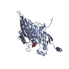 7lffC 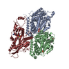 7tqxC  7tqyC  7tqzC  7tr0C  7tr1C  7tr2C M: map data used to model this data C: citing same article ( |
|---|---|
| Similar structure data | Similarity search - Function & homology  F&H Search F&H Search |
- Links
Links
- Assembly
| Deposited unit | 
|
|---|---|
| 1 |
|
- Components
-Protein , 3 types, 3 molecules ABK
| #1: Protein | Mass: 50204.445 Da / Num. of mol.: 1 / Source method: isolated from a natural source / Source: (natural)  |
|---|---|
| #2: Protein | Mass: 49999.887 Da / Num. of mol.: 1 / Source method: isolated from a natural source / Source: (natural)  |
| #3: Protein | Mass: 54869.469 Da / Num. of mol.: 1 Source method: isolated from a genetically manipulated source Source: (gene. exp.)  Candida albicans (yeast) / Gene: KIP3, orf19.7353, CAALFM_C305720CA / Production host: Candida albicans (yeast) / Gene: KIP3, orf19.7353, CAALFM_C305720CA / Production host:  |
-Non-polymers , 5 types, 5 molecules 


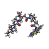


| #4: Chemical | ChemComp-GTP / |
|---|---|
| #5: Chemical | ChemComp-MG / |
| #6: Chemical | ChemComp-GDP / |
| #7: Chemical | ChemComp-SR6 / |
| #8: Chemical | ChemComp-ANP / |
-Details
| Has ligand of interest | Y |
|---|
-Experimental details
-Experiment
| Experiment | Method: ELECTRON MICROSCOPY |
|---|---|
| EM experiment | Aggregation state: FILAMENT / 3D reconstruction method: single particle reconstruction |
- Sample preparation
| Component |
| ||||||||||||||||||||||||
|---|---|---|---|---|---|---|---|---|---|---|---|---|---|---|---|---|---|---|---|---|---|---|---|---|---|
| Molecular weight | Experimental value: NO | ||||||||||||||||||||||||
| Source (natural) |
| ||||||||||||||||||||||||
| Source (recombinant) | Organism:  | ||||||||||||||||||||||||
| Buffer solution | pH: 6.8 | ||||||||||||||||||||||||
| Buffer component |
| ||||||||||||||||||||||||
| Specimen | Embedding applied: NO / Shadowing applied: NO / Staining applied: NO / Vitrification applied: YES | ||||||||||||||||||||||||
| Specimen support | Grid material: GOLD / Grid type: UltrAuFoil R2/2 | ||||||||||||||||||||||||
| Vitrification | Instrument: FEI VITROBOT MARK IV / Cryogen name: ETHANE |
- Electron microscopy imaging
| Experimental equipment |  Model: Titan Krios / Image courtesy: FEI Company |
|---|---|
| Microscopy | Model: FEI TITAN KRIOS |
| Electron gun | Electron source:  FIELD EMISSION GUN / Accelerating voltage: 300 kV / Illumination mode: FLOOD BEAM FIELD EMISSION GUN / Accelerating voltage: 300 kV / Illumination mode: FLOOD BEAM |
| Electron lens | Mode: BRIGHT FIELD / Nominal defocus max: 2670 nm / Nominal defocus min: 810 nm / Alignment procedure: COMA FREE |
| Specimen holder | Cryogen: NITROGEN / Specimen holder model: FEI TITAN KRIOS AUTOGRID HOLDER |
| Image recording | Electron dose: 64.01 e/Å2 / Detector mode: COUNTING / Film or detector model: GATAN K2 SUMMIT (4k x 4k) |
| Image scans | Movie frames/image: 50 / Used frames/image: 1-50 |
- Processing
| EM software |
| |||||||||||||||||||||||||||||||||||||||||||||||||||||||
|---|---|---|---|---|---|---|---|---|---|---|---|---|---|---|---|---|---|---|---|---|---|---|---|---|---|---|---|---|---|---|---|---|---|---|---|---|---|---|---|---|---|---|---|---|---|---|---|---|---|---|---|---|---|---|---|---|
| CTF correction | Type: PHASE FLIPPING AND AMPLITUDE CORRECTION | |||||||||||||||||||||||||||||||||||||||||||||||||||||||
| Symmetry | Point symmetry: C14 (14 fold cyclic) | |||||||||||||||||||||||||||||||||||||||||||||||||||||||
| 3D reconstruction | Resolution: 3.9 Å / Resolution method: FSC 0.143 CUT-OFF / Num. of particles: 530936 Details: The "Number of particles used" reported here is the number of asymmetric units used. Symmetry type: POINT | |||||||||||||||||||||||||||||||||||||||||||||||||||||||
| Atomic model building | Protocol: FLEXIBLE FIT / Space: REAL |
 Movie
Movie Controller


 PDBj
PDBj


