 Movie
Movie Controller


+ Open data

- Basic information
| Entry | Database: PDB / ID: 7tqb | |||||||||
|---|---|---|---|---|---|---|---|---|---|---|
| Title | Crystal structure of monoclonal S9.6 Fab bound to DNA-RNA hybrid | |||||||||
 Components Components |
| |||||||||
 Keywords Keywords | IMMUNE SYSTEM/DNA-RNA HYBRID / Antibody Fab DNA:RNA hybrid Nucleic Acid DNA:RNA hybrid binding protein / DNA-RNA HYBRID / IMMUNE SYSTEM-DNA-RNA HYBRID complex | |||||||||
| Function / homology | Immunoglobulins / Immunoglobulin-like / Sandwich / Mainly Beta / DNA / DNA (> 10) / RNA / RNA (> 10) Function and homology information Function and homology information | |||||||||
| Biological species | synthetic construct (others) | |||||||||
| Method |  X-RAY DIFFRACTION / SYNCHROTRON / MOLECULAR REPLACEMENT / molecular replacement / Resolution: 3.1 Å X-RAY DIFFRACTION / SYNCHROTRON / MOLECULAR REPLACEMENT / molecular replacement / Resolution: 3.1 Å | |||||||||
 Authors Authors | Bou-Nader, C. / Zhang, J. | |||||||||
| Funding support |  United States, 1items United States, 1items
| |||||||||
 Citation Citation | Journal: Nat Commun / Year: 2022 Title: Structural basis of R-loop recognition by the S9.6 monoclonal antibody. Authors: Bou-Nader, C. / Bothra, A. / Garboczi, D.N. / Leppla, S.H. / Zhang, J. | |||||||||
| History |
|
- Structure visualization
| Structure viewer | Molecule: MolmilJmol/JSmol |
|---|
- Downloads & links
-Download
| PDBx/mmCIF format | 7tqb.cif.gz | 208.6 KB | Display | PDBx/mmCIF format |
|---|---|---|---|---|
| PDB format | pdb7tqb.ent.gz | 159.6 KB | Display | PDB format |
| PDBx/mmJSON format | 7tqb.json.gz | Tree view | PDBx/mmJSON format | |
| Others |  Other downloads Other downloads |
-Validation report
| Arichive directory | https://data.pdbj.org/pub/pdb/validation_reports/tq/7tqbftp://data.pdbj.org/pub/pdb/validation_reports/tq/7tqb | HTTPS FTP |
|---|
-Related structure data
| Related structure data | 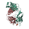 7tqaC  3tt1S S: Starting model for refinement C: citing same article ( |
|---|---|
| Similar structure data |
-Links
 PDBj
PDBj


- Assembly
| Deposited unit | 
| ||||||||
|---|---|---|---|---|---|---|---|---|---|
| 1 |
| ||||||||
| Unit cell |
|
-Components
-Antibody , 2 types, 2 molecules HL
| #1: Antibody | Mass: 24972.879 Da / Num. of mol.: 1 Source method: isolated from a genetically manipulated source Source: (gene. exp.) synthetic construct (others) / Cell line (production host): CHO / Production host:   Cricetulus griseus (Chinese hamster) Cricetulus griseus (Chinese hamster) |
|---|---|
| #2: Antibody | Mass: 24124.740 Da / Num. of mol.: 1 Source method: isolated from a genetically manipulated source Source: (gene. exp.) synthetic construct (others) / Cell line (production host): CHO / Production host: Cricetulus griseus (Chinese hamster) |
-RNA chain / DNA chain , 2 types, 2 molecules AB
| #3: RNA chain | Mass: 4094.462 Da / Num. of mol.: 1 / Source method: obtained synthetically / Source: (synth.) synthetic construct (others) |
|---|---|
| #4: DNA chain | Mass: 4000.624 Da / Num. of mol.: 1 / Source method: obtained synthetically / Source: (synth.) synthetic construct (others) |
-Non-polymers , 2 types, 37 molecules 

| #5: Chemical | ChemComp-GOL /  Mass: 92.094 Da / Num. of mol.: 1 / Source method: obtained synthetically / Formula: C3H8O3 Mass: 92.094 Da / Num. of mol.: 1 / Source method: obtained synthetically / Formula: C3H8O3 |
|---|---|
| #6: Water | ChemComp-HOH / Mass: 18.015 Da / Num. of mol.: 36 / Source method: isolated from a natural source / Formula: H2O |
-Details
| Has ligand of interest | N |
|---|---|
| Has protein modification | Y |
-Experimental details
-Experiment
| Experiment | Method: X-RAY DIFFRACTION / Number of used crystals: 1 |
|---|
- Sample preparation
| Crystal | Density Matthews: 2.18 Å3/Da / Density % sol: 43.56 % |
|---|---|
| Crystal grow | Temperature: 293 K / Method: vapor diffusion, sitting drop / Details: 30% PEG 1000 0.2 M sodium narrate |
-Data collection
| Diffraction | Mean temperature: 100 K / Serial crystal experiment: N |
|---|---|
| Diffraction source | Source: SYNCHROTRON / Site: APS / Beamline: 22-ID / Wavelength: 1 Å |
| Detector | Type: DECTRIS EIGER X 16M / Detector: PIXEL / Date: Nov 21, 2020 |
| Radiation | Protocol: SINGLE WAVELENGTH / Monochromatic (M) / Laue (L): M / Scattering type: x-ray |
| Radiation wavelength | Wavelength: 1 Å / Relative weight: 1 |
| Reflection | Resolution: 3.02→40.5 Å / Num. obs: 9509 / % possible obs: 98.7 % / Redundancy: 12.1 % / CC1/2: 0.841 / CC star: 0.956 / Net I/σ(I): 3.78 |
| Reflection shell | Resolution: 3.02→3.13 Å / Mean I/σ(I) obs: 1.55 / Num. unique obs: 925 / CC1/2: 0.393 |
-Phasing
| Phasing | Method: molecular replacement |
|---|
- Processing
| Software |
| ||||||||||||||||||||||||||||||||||||||||||||||||||||||
|---|---|---|---|---|---|---|---|---|---|---|---|---|---|---|---|---|---|---|---|---|---|---|---|---|---|---|---|---|---|---|---|---|---|---|---|---|---|---|---|---|---|---|---|---|---|---|---|---|---|---|---|---|---|---|---|
| Refinement | Method to determine structure: MOLECULAR REPLACEMENT Starting model: 3TT1 Resolution: 3.1→37.091 Å / SU ML: 0.39 / Cross valid method: THROUGHOUT / σ(F): 1.38 / Phase error: 24.65 / Stereochemistry target values: ML
| ||||||||||||||||||||||||||||||||||||||||||||||||||||||
| Solvent computation | Shrinkage radii: 0.9 Å / VDW probe radii: 1.11 Å / Solvent model: FLAT BULK SOLVENT MODEL | ||||||||||||||||||||||||||||||||||||||||||||||||||||||
| Displacement parameters | Biso max: 105.3 Å2 / Biso mean: 30.6873 Å2 / Biso min: 12.31 Å2 | ||||||||||||||||||||||||||||||||||||||||||||||||||||||
| Refinement step | Cycle: final / Resolution: 3.1→37.091 Å
| ||||||||||||||||||||||||||||||||||||||||||||||||||||||
| LS refinement shell | Refine-ID: X-RAY DIFFRACTION / Rfactor Rfree error: 0
| ||||||||||||||||||||||||||||||||||||||||||||||||||||||
| Refinement TLS params. | Method: refined / Origin x: 12.6181 Å / Origin y: -28.8717 Å / Origin z: 12.3193 Å
| ||||||||||||||||||||||||||||||||||||||||||||||||||||||
| Refinement TLS group |
|
