 Movie
Movie Controller


+ Open data

- Basic information
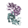
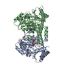
| Entry | Database: PDB / ID: 7tlp | ||||||
|---|---|---|---|---|---|---|---|
| Title | Structure of Atopobium parvulum SufS K235R | ||||||
 Components Components | Cysteine desulfurase | ||||||
 Keywords Keywords | TRANSFERASE / Cysteine desulferase | ||||||
| Function / homology |  Function and homology information Function and homology informationcysteine desulfurase / cysteine desulfurase activity / : / pyridoxal phosphate binding Similarity search - Function | ||||||
| Biological species |  Lancefieldella parvula (bacteria) Lancefieldella parvula (bacteria) | ||||||
| Method |  X-RAY DIFFRACTION / MOLECULAR REPLACEMENT / Resolution: 2.99 Å X-RAY DIFFRACTION / MOLECULAR REPLACEMENT / Resolution: 2.99 Å | ||||||
 Authors Authors | Karunakaran, G. / Couture, J.F. | ||||||
| Funding support |  Canada, 1items Canada, 1items
| ||||||
 Citation Citation | Journal: Febs Lett. / Year: 2022 Title: Structural analysis of Atopobium parvulum SufS cysteine desulfurase linked to Crohn's disease. Authors: Karunakaran, G. / Yang, Y. / Tremblay, V. / Ning, Z. / Martin, J. / Belaouad, A. / Figeys, D. / Brunzelle, J.S. / Giguere, P.M. / Stintzi, A. / Couture, J.F. | ||||||
| History |
|
- Structure visualization
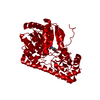
| Structure viewer | Molecule: MolmilJmol/JSmol |
|---|
- Downloads & links
-Download
| PDBx/mmCIF format | 7tlp.cif.gz | 965.9 KB | Display | PDBx/mmCIF format |
|---|---|---|---|---|
| PDB format | pdb7tlp.ent.gz | 656.9 KB | Display | PDB format |
| PDBx/mmJSON format | 7tlp.json.gz | Tree view | PDBx/mmJSON format | |
| Others |  Other downloads Other downloads |
-Validation report
| Arichive directory | https://data.pdbj.org/pub/pdb/validation_reports/tl/7tlpftp://data.pdbj.org/pub/pdb/validation_reports/tl/7tlp | HTTPS FTP |
|---|
-Related structure data
| Related structure data |  7tlmC  7tlqC  7tlrC  5j8qS S: Starting model for refinement C: citing same article ( |
|---|---|
| Similar structure data |

-Links
 PDBj
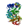
PDBj- Assembly
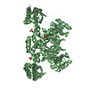
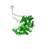
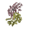
| Deposited unit | 
| |||||||||||||||||||||||||||||||||||||||||||||||||||||||||||||||||||||||||||||||||||||||||||||||||||||||||||||||||||||||||||
|---|---|---|---|---|---|---|---|---|---|---|---|---|---|---|---|---|---|---|---|---|---|---|---|---|---|---|---|---|---|---|---|---|---|---|---|---|---|---|---|---|---|---|---|---|---|---|---|---|---|---|---|---|---|---|---|---|---|---|---|---|---|---|---|---|---|---|---|---|---|---|---|---|---|---|---|---|---|---|---|---|---|---|---|---|---|---|---|---|---|---|---|---|---|---|---|---|---|---|---|---|---|---|---|---|---|---|---|---|---|---|---|---|---|---|---|---|---|---|---|---|---|---|---|---|
| 1 | 
| |||||||||||||||||||||||||||||||||||||||||||||||||||||||||||||||||||||||||||||||||||||||||||||||||||||||||||||||||||||||||||
| 2 | 
| |||||||||||||||||||||||||||||||||||||||||||||||||||||||||||||||||||||||||||||||||||||||||||||||||||||||||||||||||||||||||||
| 3 | 
| |||||||||||||||||||||||||||||||||||||||||||||||||||||||||||||||||||||||||||||||||||||||||||||||||||||||||||||||||||||||||||
| 4 | 
| |||||||||||||||||||||||||||||||||||||||||||||||||||||||||||||||||||||||||||||||||||||||||||||||||||||||||||||||||||||||||||
| Unit cell |
| |||||||||||||||||||||||||||||||||||||||||||||||||||||||||||||||||||||||||||||||||||||||||||||||||||||||||||||||||||||||||||
| Noncrystallographic symmetry (NCS) | NCS domain:
NCS domain segments:
|