 Movie
Movie Controller

[English] 日本語
 Yorodumi
- PDB-7t92: Structure of the peroxisomal retro-translocon formed by a heterot... -
+ Open data

- Basic information
| Entry | Database: PDB / ID: 7t92 | ||||||
|---|---|---|---|---|---|---|---|
| Title | Structure of the peroxisomal retro-translocon formed by a heterotrimeric ubiquitin ligase complex | ||||||
 Components Components |
| ||||||
 Keywords Keywords | TRANSLOCASE / peroxisome / retro-translocon / ubiquitin ligase | ||||||
| Function / homology |  Function and homology information Function and homology informationperoxisomal importomer complex / RING-type E3 ubiquitin transferase (cysteine targeting) / protein import into peroxisome matrix, receptor recycling / peroxisomal membrane / protein monoubiquitination / RING-type E3 ubiquitin transferase / ubiquitin-protein transferase activity / ubiquitin protein ligase activity / protein ubiquitination / zinc ion binding Similarity search - Function | ||||||
| Biological species |  Thermothelomyces thermophilus ATCC 42464 (fungus) Thermothelomyces thermophilus ATCC 42464 (fungus)synthetic construct (others) | ||||||
| Method | ELECTRON MICROSCOPY / single particle reconstruction / cryo EM / Resolution: 3.1 Å | ||||||
 Authors Authors | Peiqiang, F. / Tom, R. | ||||||
| Funding support |  United States, 1items United States, 1items
| ||||||
 Citation Citation | Journal: Nature / Year: 2022 Title: A peroxisomal ubiquitin ligase complex forms a retrotranslocation channel. Authors: Peiqiang Feng / Xudong Wu / Satchal K Erramilli / Joao A Paulo / Pawel Knejski / Steven P Gygi / Anthony A Kossiakoff / Tom A Rapoport / Abstract: Peroxisomes are ubiquitous organelles that house various metabolic reactions and are essential for human health. Luminal peroxisomal proteins are imported from the cytosol by mobile receptors, which ...Peroxisomes are ubiquitous organelles that house various metabolic reactions and are essential for human health. Luminal peroxisomal proteins are imported from the cytosol by mobile receptors, which then recycle back to the cytosol by a poorly understood process. Recycling requires receptor modification by a membrane-embedded ubiquitin ligase complex comprising three RING finger domain-containing proteins (Pex2, Pex10 and Pex12). Here we report a cryo-electron microscopy structure of the ligase complex, which together with biochemical and in vivo experiments reveals its function as a retrotranslocation channel for peroxisomal import receptors. Each subunit of the complex contributes five transmembrane segments that co-assemble into an open channel. The three ring finger domains form a cytosolic tower, with ring finger 2 (RF2) positioned above the channel pore. We propose that the N terminus of a recycling receptor is inserted from the peroxisomal lumen into the pore and monoubiquitylated by RF2 to enable extraction into the cytosol. If recycling is compromised, receptors are polyubiquitylated by the concerted action of RF10 and RF12 and degraded. This polyubiquitylation pathway also maintains the homeostasis of other peroxisomal import factors. Our results clarify a crucial step during peroxisomal protein import and reveal why mutations in the ligase complex cause human disease. | ||||||
| History |
|
- Structure visualization
| Structure viewer | Molecule: MolmilJmol/JSmol |
|---|
- Downloads & links
-Download
| PDBx/mmCIF format | 7t92.cif.gz | 430.6 KB | Display | PDBx/mmCIF format |
|---|---|---|---|---|
| PDB format | pdb7t92.ent.gz | 348.7 KB | Display | PDB format |
| PDBx/mmJSON format | 7t92.json.gz | Tree view | PDBx/mmJSON format | |
| Others |  Other downloads Other downloads |
-Validation report
| Arichive directory | https://data.pdbj.org/pub/pdb/validation_reports/t9/7t92ftp://data.pdbj.org/pub/pdb/validation_reports/t9/7t92 | HTTPS FTP |
|---|
-Related structure data
| Related structure data |  25750MC  7t9xC M: map data used to model this data C: citing same article ( |
|---|---|
| Similar structure data |
-Links
 PDBj
PDBj


- Assembly
| Deposited unit | 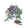
|
|---|---|
| 1 |
|
-Components
-Protein , 3 types, 3 molecules BAC
| #1: Protein | Mass: 49369.703 Da / Num. of mol.: 1 Source method: isolated from a genetically manipulated source Source: (gene. exp.) Thermothelomyces thermophilus ATCC 42464 (fungus)Strain: ATCC 42464 / BCRC 31852 / DSM 1799 / Gene: MYCTH_2053677 / Production host: Komagataella pastoris (fungus) / References: UniProt: G2Q5N0 |
|---|---|
| #2: Protein | Mass: 38493.715 Da / Num. of mol.: 1 Source method: isolated from a genetically manipulated source Source: (gene. exp.) Thermothelomyces thermophilus ATCC 42464 (fungus)Strain: ATCC 42464 / BCRC 31852 / DSM 1799 / Gene: MYCTH_2294472 / Production host: Komagataella pastoris (fungus) / References: UniProt: G2Q1C9 |
| #3: Protein | Mass: 48952.234 Da / Num. of mol.: 1 Source method: isolated from a genetically manipulated source Source: (gene. exp.) Thermothelomyces thermophilus ATCC 42464 (fungus)Strain: ATCC 42464 / BCRC 31852 / DSM 1799 / Gene: MYCTH_2294231 / Production host: Komagataella pastoris (fungus) / References: UniProt: G2Q0E2 |
-Antibody , 2 types, 2 molecules HL
| #4: Antibody | Mass: 12518.806 Da / Num. of mol.: 1 Source method: isolated from a genetically manipulated source Source: (gene. exp.) synthetic construct (others) / Production host:  |
|---|---|
| #5: Antibody | Mass: 11135.375 Da / Num. of mol.: 1 Source method: isolated from a genetically manipulated source Source: (gene. exp.) synthetic construct (others) / Production host: |
-Non-polymers , 3 types, 25 molecules 

| #6: Chemical | ChemComp-ZN /  Mass: 65.409 Da / Num. of mol.: 5 / Source method: obtained synthetically / Formula: Zn Mass: 65.409 Da / Num. of mol.: 5 / Source method: obtained synthetically / Formula: Zn#7: Chemical | ChemComp-LBN /  Mass: 760.076 Da / Num. of mol.: 15 / Source method: obtained synthetically / Formula: C42H82NO8P / Comment: phospholipid*YM Mass: 760.076 Da / Num. of mol.: 15 / Source method: obtained synthetically / Formula: C42H82NO8P / Comment: phospholipid*YM#8: Chemical | ChemComp-CLR /  Mass: 386.654 Da / Num. of mol.: 5 / Source method: obtained synthetically / Formula: C27H46O Mass: 386.654 Da / Num. of mol.: 5 / Source method: obtained synthetically / Formula: C27H46O |
|---|
-Details
| Has ligand of interest | N |
|---|
-Experimental details
-Experiment
| Experiment | Method: ELECTRON MICROSCOPY |
|---|---|
| EM experiment | Aggregation state: PARTICLE / 3D reconstruction method: single particle reconstruction |
- Sample preparation
| Component |
| ||||||||||||||||||||||||
|---|---|---|---|---|---|---|---|---|---|---|---|---|---|---|---|---|---|---|---|---|---|---|---|---|---|
| Molecular weight | Experimental value: NO | ||||||||||||||||||||||||
| Source (natural) |
| ||||||||||||||||||||||||
| Source (recombinant) |
| ||||||||||||||||||||||||
| Buffer solution | pH: 7.4 | ||||||||||||||||||||||||
| Specimen | Embedding applied: NO / Shadowing applied: NO / Staining applied: NO / Vitrification applied: YES / Details: This sample was monodisperse. | ||||||||||||||||||||||||
| Vitrification | Instrument: FEI VITROBOT MARK I / Cryogen name: ETHANE / Humidity: 100 % / Chamber temperature: 4 K |
- Electron microscopy imaging
| Experimental equipment |  Model: Titan Krios / Image courtesy: FEI Company |
|---|---|
| Microscopy | Model: FEI TITAN KRIOS |
| Electron gun | Electron source:  FIELD EMISSION GUN / Accelerating voltage: 300 kV / Illumination mode: FLOOD BEAM FIELD EMISSION GUN / Accelerating voltage: 300 kV / Illumination mode: FLOOD BEAM |
| Electron lens | Mode: BRIGHT FIELD / Nominal defocus max: 2500 nm / Nominal defocus min: 1000 nm |
| Image recording | Electron dose: 52 e/Å2 / Detector mode: COUNTING / Film or detector model: GATAN K2 SUMMIT (4k x 4k) |
- Processing
| Software |
| ||||||||||||||||||||||||
|---|---|---|---|---|---|---|---|---|---|---|---|---|---|---|---|---|---|---|---|---|---|---|---|---|---|
| CTF correction | Type: PHASE FLIPPING AND AMPLITUDE CORRECTION | ||||||||||||||||||||||||
| 3D reconstruction | Resolution: 3.1 Å / Resolution method: FSC 0.143 CUT-OFF / Num. of particles: 121644 / Symmetry type: POINT | ||||||||||||||||||||||||
| Refinement | Highest resolution: 3.1 Å / Cross valid method: NONE | ||||||||||||||||||||||||
| Refine LS restraints |
|
