 Movie
Movie Controller

[English] 日本語
 Yorodumi
- PDB-7t6i: Crystal structure of HLA-DP1 in complex with pp65 peptide in reve... -
+ Open data

- Basic information
| Entry | Database: PDB / ID: 7t6i | ||||||
|---|---|---|---|---|---|---|---|
| Title | Crystal structure of HLA-DP1 in complex with pp65 peptide in reverse orientation | ||||||
 Components Components |
| ||||||
 Keywords Keywords | IMMUNE SYSTEM / Antigen presentation | ||||||
| Function / homology |  Function and homology information Function and homology informationviral tegument / antigen processing and presentation of peptide or polysaccharide antigen via MHC class II / MHC class II protein complex / Immunoregulatory interactions between a Lymphoid and a non-Lymphoid cell / symbiont-mediated suppression of host cytoplasmic pattern recognition receptor signaling pathway via inhibition of IRF3 activity / adaptive immune response / host cell cytoplasm / membrane => GO:0016020 / endosome membrane / lysosomal membrane ...viral tegument / antigen processing and presentation of peptide or polysaccharide antigen via MHC class II / MHC class II protein complex / Immunoregulatory interactions between a Lymphoid and a non-Lymphoid cell / symbiont-mediated suppression of host cytoplasmic pattern recognition receptor signaling pathway via inhibition of IRF3 activity / adaptive immune response / host cell cytoplasm / membrane => GO:0016020 / endosome membrane / lysosomal membrane / virus-mediated perturbation of host defense response / host cell nucleus Similarity search - Function | ||||||
| Biological species |  Homo sapiens (human) Homo sapiens (human)  Human cytomegalovirus Human cytomegalovirus | ||||||
| Method |  X-RAY DIFFRACTION / SYNCHROTRON / MOLECULAR REPLACEMENT / Resolution: 2.3 Å X-RAY DIFFRACTION / SYNCHROTRON / MOLECULAR REPLACEMENT / Resolution: 2.3 Å | ||||||
 Authors Authors | Lim, J.J. / Reid, H. / Rossjohn, J. | ||||||
| Funding support |  Australia, 1items Australia, 1items
| ||||||
 Citation Citation | Journal: Proc.Natl.Acad.Sci.USA / Year: 2022 Title: Human T cells recognize HLA-DP-bound peptides in two orientations. Authors: Klobuch, S. / Lim, J.J. / van Balen, P. / Kester, M.G.D. / de Klerk, W. / de Ru, A.H. / Pothast, C.R. / Jedema, I. / Drijfhout, J.W. / Rossjohn, J. / Reid, H.H. / van Veelen, P.A. / ...Authors: Klobuch, S. / Lim, J.J. / van Balen, P. / Kester, M.G.D. / de Klerk, W. / de Ru, A.H. / Pothast, C.R. / Jedema, I. / Drijfhout, J.W. / Rossjohn, J. / Reid, H.H. / van Veelen, P.A. / Falkenburg, J.H.F. / Heemskerk, M.H.M. | ||||||
| History |
|
- Structure visualization
| Structure viewer | Molecule: MolmilJmol/JSmol |
|---|
- Downloads & links
-Download
| PDBx/mmCIF format | 7t6i.cif.gz | 117.3 KB | Display | PDBx/mmCIF format |
|---|---|---|---|---|
| PDB format | pdb7t6i.ent.gz | 71.4 KB | Display | PDB format |
| PDBx/mmJSON format | 7t6i.json.gz | Tree view | PDBx/mmJSON format | |
| Others |  Other downloads Other downloads |
-Validation report
| Summary document | 7t6i_validation.pdf.gz | 478.5 KB | Display | wwPDB validaton report |
|---|---|---|---|---|
| Full document | 7t6i_full_validation.pdf.gz | 479.2 KB | Display | |
| Data in XML | 7t6i_validation.xml.gz | 17.5 KB | Display | |
| Data in CIF | 7t6i_validation.cif.gz | 24.4 KB | Display | |
| Arichive directory | https://data.pdbj.org/pub/pdb/validation_reports/t6/7t6iftp://data.pdbj.org/pub/pdb/validation_reports/t6/7t6i | HTTPS FTP |
-Related structure data
| Related structure data |  3wexS S: Starting model for refinement |
|---|---|
| Similar structure data |
-Links
 PDBj
PDBj


- Assembly
| Deposited unit | 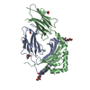
| ||||||||||||
|---|---|---|---|---|---|---|---|---|---|---|---|---|---|
| 1 |
| ||||||||||||
| Unit cell |
|
-Components
-Protein , 2 types, 2 molecules AB
| #1: Protein | Mass: 21020.340 Da / Num. of mol.: 1 Source method: isolated from a genetically manipulated source Source: (gene. exp.) Homo sapiens (human) / Gene: HLA-DPA1Production host: Insect expression vector pBlueBacmsGCA1His (others) References: UniProt: Q95HB9 |
|---|---|
| #2: Protein | Mass: 22173.719 Da / Num. of mol.: 1 Source method: isolated from a genetically manipulated source Source: (gene. exp.) Homo sapiens (human) / Gene: HLA-DPB1, DPB1Production host: Insect expression vector pBlueBacmsGCB1His (others) References: UniProt: S6B6U4 |
-Protein/peptide / Sugars , 2 types, 4 molecules C
| #3: Protein/peptide | Mass: 1853.148 Da / Num. of mol.: 1 / Fragment: residues 142-158 / Source method: obtained synthetically / Source: (synth.)  Human cytomegalovirus (strain AD169) / References: UniProt: P06725 Human cytomegalovirus (strain AD169) / References: UniProt: P06725 |
|---|---|
| #4: Sugar |  Type: D-saccharide, beta linking / Mass: 221.208 Da / Num. of mol.: 3 / Source method: obtained synthetically / Formula: C8H15NO6 Type: D-saccharide, beta linking / Mass: 221.208 Da / Num. of mol.: 3 / Source method: obtained synthetically / Formula: C8H15NO6 |
-Non-polymers , 3 types, 193 molecules 


| #5: Chemical | ChemComp-GLY /  Type: peptide linking / Mass: 75.067 Da / Num. of mol.: 1 / Source method: obtained synthetically / Formula: C2H5NO2 Type: peptide linking / Mass: 75.067 Da / Num. of mol.: 1 / Source method: obtained synthetically / Formula: C2H5NO2 |
|---|---|
| #6: Chemical | ChemComp-EDO /  Mass: 62.068 Da / Num. of mol.: 1 / Source method: obtained synthetically / Formula: C2H6O2 Mass: 62.068 Da / Num. of mol.: 1 / Source method: obtained synthetically / Formula: C2H6O2 |
| #7: Water | ChemComp-HOH / Mass: 18.015 Da / Num. of mol.: 191 / Source method: isolated from a natural source / Formula: H2O |
-Details
| Has ligand of interest | N |
|---|
-Experimental details
-Experiment
| Experiment | Method: X-RAY DIFFRACTION / Number of used crystals: 1 |
|---|
- Sample preparation
| Crystal | Density Matthews: 2.83 Å3/Da / Density % sol: 56.48 % |
|---|---|
| Crystal grow | Temperature: 293.15 K / Method: vapor diffusion, sitting drop / pH: 6.5 / Details: 0.1 M Sodium Cacodylate pH 6.5 25% w/v PEG 4000 |
-Data collection
| Diffraction | Mean temperature: 100 K / Serial crystal experiment: N |
|---|---|
| Diffraction source | Source: SYNCHROTRON / Site: Australian Synchrotron / Beamline: MX2 / Wavelength: 0.9537 Å |
| Detector | Type: DECTRIS EIGER X 16M / Detector: PIXEL / Date: Jul 30, 2021 |
| Radiation | Protocol: SINGLE WAVELENGTH / Monochromatic (M) / Laue (L): M / Scattering type: x-ray |
| Radiation wavelength | Wavelength: 0.9537 Å / Relative weight: 1 |
| Reflection | Resolution: 2.3→45.04 Å / Num. obs: 22454 / % possible obs: 99.7 % / Redundancy: 5.8 % / Biso Wilson estimate: 33.02 Å2 / CC1/2: 0.997 / Net I/σ(I): 12.7 |
| Reflection shell | Resolution: 2.3→2.38 Å / Rmerge(I) obs: 0.255 / Mean I/σ(I) obs: 5.9 / Num. unique obs: 2175 / CC1/2: 0.956 |
- Processing
| Software |
| |||||||||||||||||||||||||||||||||||||||||||||||||||||||||||||||
|---|---|---|---|---|---|---|---|---|---|---|---|---|---|---|---|---|---|---|---|---|---|---|---|---|---|---|---|---|---|---|---|---|---|---|---|---|---|---|---|---|---|---|---|---|---|---|---|---|---|---|---|---|---|---|---|---|---|---|---|---|---|---|---|---|
| Refinement | Method to determine structure: MOLECULAR REPLACEMENT Starting model: 3WEX Resolution: 2.3→43.91 Å / SU ML: 0.2547 / Cross valid method: FREE R-VALUE / σ(F): 1.39 / Phase error: 22.8108 Stereochemistry target values: GeoStd + Monomer Library + CDL v1.2
| |||||||||||||||||||||||||||||||||||||||||||||||||||||||||||||||
| Solvent computation | Shrinkage radii: 0.9 Å / VDW probe radii: 1.11 Å / Solvent model: FLAT BULK SOLVENT MODEL | |||||||||||||||||||||||||||||||||||||||||||||||||||||||||||||||
| Displacement parameters | Biso mean: 37.07 Å2 | |||||||||||||||||||||||||||||||||||||||||||||||||||||||||||||||
| Refinement step | Cycle: LAST / Resolution: 2.3→43.91 Å
| |||||||||||||||||||||||||||||||||||||||||||||||||||||||||||||||
| Refine LS restraints |
| |||||||||||||||||||||||||||||||||||||||||||||||||||||||||||||||
| LS refinement shell |
|
