 Movie
Movie Controller


+ Open data

- Basic information
| Entry | Database: PDB / ID: 7t5w | ||||||
|---|---|---|---|---|---|---|---|
| Title | Structure of E. coli CapH C-terminal domain | ||||||
 Components Components | Helix-turn-helix domain-containing protein | ||||||
 Keywords Keywords | DNA BINDING PROTEIN / helix turn helix / HTH / DdrO | ||||||
| Function / homology |  Function and homology information Function and homology information | ||||||
| Biological species |  | ||||||
| Method |  X-RAY DIFFRACTION / SYNCHROTRON / MOLECULAR REPLACEMENT / Resolution: 1.75 Å X-RAY DIFFRACTION / SYNCHROTRON / MOLECULAR REPLACEMENT / Resolution: 1.75 Å | ||||||
 Authors Authors | Lau, R.K. / Corbett, K.D. | ||||||
| Funding support |  United States, 1items United States, 1items
| ||||||
 Citation Citation | Journal: Embo J. / Year: 2022 Title: A conserved signaling pathway activates bacterial CBASS immune signaling in response to DNA damage. Authors: Lau, R.K. / Enustun, E. / Gu, Y. / Nguyen, J.V. / Corbett, K.D. | ||||||
| History |
|
- Structure visualization
| Structure viewer | Molecule: MolmilJmol/JSmol |
|---|
- Downloads & links
-Download
| PDBx/mmCIF format | 7t5w.cif.gz | 121.9 KB | Display | PDBx/mmCIF format |
|---|---|---|---|---|
| PDB format | pdb7t5w.ent.gz | 79.5 KB | Display | PDB format |
| PDBx/mmJSON format | 7t5w.json.gz | Tree view | PDBx/mmJSON format | |
| Others |  Other downloads Other downloads |
-Validation report
| Arichive directory | https://data.pdbj.org/pub/pdb/validation_reports/t5/7t5wftp://data.pdbj.org/pub/pdb/validation_reports/t5/7t5w | HTTPS FTP |
|---|
-Related structure data
| Related structure data | 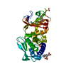 7t5tC 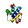 7t5uC  7t5vSC S: Starting model for refinement C: citing same article ( |
|---|---|
| Similar structure data | |
| Experimental dataset #1 | Data reference: 10.15785/SBGRID/867 / Data set type: diffraction image data / Metadata reference: 10.15785/SBGRID/867 |
-Links
 PDBj
PDBj


- Assembly
| Deposited unit | 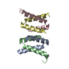
| ||||||||||||
|---|---|---|---|---|---|---|---|---|---|---|---|---|---|
| 1 |
| ||||||||||||
| Unit cell |
|
-Components
| #1: Protein/peptide | Mass: 5277.855 Da / Num. of mol.: 4 / Fragment: C-terminal residues 67-107 Source method: isolated from a genetically manipulated source Source: (gene. exp.) Gene: nadR_1, BHS87_27750, D9K17_19515, GRQ19_13110, HV109_14215, NCTC13216_00230 Production host: #2: Water | ChemComp-HOH / |  Mass: 18.015 Da / Num. of mol.: 60 / Source method: isolated from a natural source / Formula: H2O Mass: 18.015 Da / Num. of mol.: 60 / Source method: isolated from a natural source / Formula: H2O |
|---|
-Experimental details
-Experiment
| Experiment | Method: X-RAY DIFFRACTION / Number of used crystals: 1 |
|---|
- Sample preparation
| Crystal | Density Matthews: 1.9 Å3/Da / Density % sol: 35.26 % |
|---|---|
| Crystal grow | Temperature: 293 K / Method: vapor diffusion, hanging drop / Details: 0.1 M Sodium Citrate pH 3.0, 1.6 M LiSO4 |
-Data collection
| Diffraction | Mean temperature: 100 K / Serial crystal experiment: N |
|---|---|
| Diffraction source | Source: SYNCHROTRON / Site: ALS / Beamline: 5.0.2 / Wavelength: 1.00004 Å |
| Detector | Type: DECTRIS PILATUS3 6M / Detector: PIXEL / Date: May 14, 2021 |
| Radiation | Protocol: SINGLE WAVELENGTH / Monochromatic (M) / Laue (L): M / Scattering type: x-ray |
| Radiation wavelength | Wavelength: 1.00004 Å / Relative weight: 1 |
| Reflection | Resolution: 1.75→100 Å / Num. obs: 15936 / % possible obs: 99.5 % / Redundancy: 3.2 % / Biso Wilson estimate: 31.13 Å2 / CC1/2: 1 / Rpim(I) all: 0.039 / Rrim(I) all: 0.07 / Net I/σ(I): 26.2 |
| Reflection shell | Resolution: 1.75→1.78 Å / Mean I/σ(I) obs: 1.1 / Num. unique obs: 768 / CC1/2: 0.631 / Rpim(I) all: 0.45 / Rrim(I) all: 0.784 |
- Processing
| Software |
| |||||||||||||||||||||||||||||||||||||||||||||||||||||||||||||||||||||||||||
|---|---|---|---|---|---|---|---|---|---|---|---|---|---|---|---|---|---|---|---|---|---|---|---|---|---|---|---|---|---|---|---|---|---|---|---|---|---|---|---|---|---|---|---|---|---|---|---|---|---|---|---|---|---|---|---|---|---|---|---|---|---|---|---|---|---|---|---|---|---|---|---|---|---|---|---|---|
| Refinement | Method to determine structure: MOLECULAR REPLACEMENT Starting model: 7T5V Resolution: 1.75→45.72 Å / SU ML: 0.2478 / Cross valid method: FREE R-VALUE / σ(F): 1.39 / Phase error: 28.585 Stereochemistry target values: GeoStd + Monomer Library + CDL v1.2
| |||||||||||||||||||||||||||||||||||||||||||||||||||||||||||||||||||||||||||
| Solvent computation | Shrinkage radii: 0.9 Å / VDW probe radii: 1.11 Å / Solvent model: FLAT BULK SOLVENT MODEL | |||||||||||||||||||||||||||||||||||||||||||||||||||||||||||||||||||||||||||
| Displacement parameters | Biso mean: 37.08 Å2 | |||||||||||||||||||||||||||||||||||||||||||||||||||||||||||||||||||||||||||
| Refinement step | Cycle: LAST / Resolution: 1.75→45.72 Å
| |||||||||||||||||||||||||||||||||||||||||||||||||||||||||||||||||||||||||||
| Refine LS restraints |
| |||||||||||||||||||||||||||||||||||||||||||||||||||||||||||||||||||||||||||
| LS refinement shell |
| |||||||||||||||||||||||||||||||||||||||||||||||||||||||||||||||||||||||||||
| Refinement TLS params. | Method: refined / Refine-ID: X-RAY DIFFRACTION
| |||||||||||||||||||||||||||||||||||||||||||||||||||||||||||||||||||||||||||
| Refinement TLS group | Refine-ID: X-RAY DIFFRACTION
|
