 Movie
Movie Controller

[English] 日本語
 Yorodumi
- PDB-7t4z: Crystal structure of the molybdate-binding periplasmic protein Mo... -
+ Open data

- Basic information
| Entry | Database: PDB / ID: 7t4z | ||||||||||||||||||||||||
|---|---|---|---|---|---|---|---|---|---|---|---|---|---|---|---|---|---|---|---|---|---|---|---|---|---|
| Title | Crystal structure of the molybdate-binding periplasmic protein ModA from the bacteria Pseudomonsa aeruginosa in ligand-free form | ||||||||||||||||||||||||
 Components Components | Molybdate-binding periplasmic protein | ||||||||||||||||||||||||
 Keywords Keywords | METAL BINDING PROTEIN / Molybdate-binding periplasmic protein | ||||||||||||||||||||||||
| Function / homology | AMMONIUM ION Function and homology information Function and homology information | ||||||||||||||||||||||||
| Biological species |  Pseudomonas aeruginosa PA1 (bacteria) Pseudomonas aeruginosa PA1 (bacteria) | ||||||||||||||||||||||||
| Method |  X-RAY DIFFRACTION / SYNCHROTRON / MOLECULAR REPLACEMENT / Resolution: 1.78 Å X-RAY DIFFRACTION / SYNCHROTRON / MOLECULAR REPLACEMENT / Resolution: 1.78 Å | ||||||||||||||||||||||||
 Authors Authors | Ngu, D.H.Y. / Luo, Z. / Lim, B.Y.J. / Kobe, B. | ||||||||||||||||||||||||
| Funding support |  Australia, 7items Australia, 7items
| ||||||||||||||||||||||||
 Citation Citation | Journal: Front Microbiol / Year: 2022 Title: The Impact of Chromate on Pseudomonas aeruginosa Molybdenum Homeostasis. Authors: Maunders, E.A. / Ngu, D.H.Y. / Ganio, K. / Hossain, S.I. / Lim, B.Y.J. / Leeming, M.G. / Luo, Z. / Tan, A. / Deplazes, E. / Kobe, B. / McDevitt, C.A. | ||||||||||||||||||||||||
| History |
|
- Structure visualization
| Structure viewer | Molecule: MolmilJmol/JSmol |
|---|
- Downloads & links
-Download
| PDBx/mmCIF format | 7t4z.cif.gz | 297.4 KB | Display | PDBx/mmCIF format |
|---|---|---|---|---|
| PDB format | pdb7t4z.ent.gz | 213.1 KB | Display | PDB format |
| PDBx/mmJSON format | 7t4z.json.gz | Tree view | PDBx/mmJSON format | |
| Others |  Other downloads Other downloads |
-Validation report
| Arichive directory | https://data.pdbj.org/pub/pdb/validation_reports/t4/7t4zftp://data.pdbj.org/pub/pdb/validation_reports/t4/7t4z | HTTPS FTP |
|---|
-Related structure data
| Related structure data |  7t50C  7t51C  7t5aC  1atgS S: Starting model for refinement C: citing same article ( |
|---|---|
| Similar structure data |
-Links
 PDBj
PDBj
- Assembly
| Deposited unit | 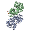
| ||||||||||||||||||||||||||||||||||||||||||||||||||||||||||
|---|---|---|---|---|---|---|---|---|---|---|---|---|---|---|---|---|---|---|---|---|---|---|---|---|---|---|---|---|---|---|---|---|---|---|---|---|---|---|---|---|---|---|---|---|---|---|---|---|---|---|---|---|---|---|---|---|---|---|---|
| 1 | 
| ||||||||||||||||||||||||||||||||||||||||||||||||||||||||||
| 2 | 
| ||||||||||||||||||||||||||||||||||||||||||||||||||||||||||
| Unit cell |
| ||||||||||||||||||||||||||||||||||||||||||||||||||||||||||
| Components on special symmetry positions |
| ||||||||||||||||||||||||||||||||||||||||||||||||||||||||||
| Noncrystallographic symmetry (NCS) | NCS domain:
NCS domain segments:
NCS oper: (Code: givenMatrix: (0.529418366487, 0.785485412688, -0.320513431356), (0.750580081135, -0.609770766846, -0.254576420164), (-0.395405785276, -0.105793564803, -0.912393548101)Vector: -13. ...NCS oper: (Code: given Matrix: (0.529418366487, 0.785485412688, -0.320513431356), Vector: |
-Components
| #1: Protein | Mass: 24379.453 Da / Num. of mol.: 2 Source method: isolated from a genetically manipulated source Source: (gene. exp.) Pseudomonas aeruginosa PA1 (bacteria) / Strain: Laboratory strain PAO1 / Gene: modA / Plasmid: pMCSG7 / Production host: #2: Chemical | ChemComp-GOL / |   Mass: 92.094 Da / Num. of mol.: 1 / Source method: obtained synthetically / Formula: C3H8O3 Mass: 92.094 Da / Num. of mol.: 1 / Source method: obtained synthetically / Formula: C3H8O3#3: Chemical | ChemComp-NH4 / |   Mass: 18.038 Da / Num. of mol.: 1 / Source method: obtained synthetically / Formula: H4N Mass: 18.038 Da / Num. of mol.: 1 / Source method: obtained synthetically / Formula: H4N#4: Chemical |   Mass: 96.063 Da / Num. of mol.: 2 / Source method: obtained synthetically / Formula: SO4 Mass: 96.063 Da / Num. of mol.: 2 / Source method: obtained synthetically / Formula: SO4#5: Water | ChemComp-HOH / |  Mass: 18.015 Da / Num. of mol.: 278 / Source method: isolated from a natural source / Formula: H2O Mass: 18.015 Da / Num. of mol.: 278 / Source method: isolated from a natural source / Formula: H2OHas ligand of interest | N | |
|---|
-Experimental details
-Experiment
| Experiment | Method: X-RAY DIFFRACTION / Number of used crystals: 1 |
|---|
- Sample preparation
| Crystal | Density Matthews: 2.23 Å3/Da / Density % sol: 45 % |
|---|---|
| Crystal grow | Temperature: 293.15 K / Method: vapor diffusion, hanging drop / pH: 3.5 Details: 0.1 M sodium citrate pH 3.5, 2.0 M ammonium sulfate, 25% v/v glycerol |
-Data collection
| Diffraction | Mean temperature: 100 K / Serial crystal experiment: N |
|---|---|
| Diffraction source | Source: SYNCHROTRON / Site: Australian Synchrotron / Beamline: MX2 / Wavelength: 0.954 Å |
| Detector | Type: DECTRIS EIGER X 16M / Detector: PIXEL / Date: Aug 15, 2019 |
| Radiation | Protocol: SINGLE WAVELENGTH / Monochromatic (M) / Laue (L): M / Scattering type: x-ray |
| Radiation wavelength | Wavelength: 0.954 Å / Relative weight: 1 |
| Reflection | Resolution: 1.78→46.68 Å / Num. obs: 44640 / % possible obs: 99.9 % / Redundancy: 6.8 % / Biso Wilson estimate: 21.64 Å2 / CC1/2: 0.999 / CC star: 1 / Rmerge(I) obs: 0.067 / Rpim(I) all: 0.028 / Rrim(I) all: 0.073 / Net I/σ(I): 16.7 |
| Reflection shell | Resolution: 1.78→1.82 Å / Rmerge(I) obs: 0.518 / Mean I/σ(I) obs: 3.5 / Num. unique obs: 2473 / CC1/2: 0.896 / CC star: 0.972 / Rpim(I) all: 0.214 / Rrim(I) all: 0.562 / % possible all: 98.2 |
- Processing
| Software |
| |||||||||||||||||||||||||||||||||||||||||||||||||||||||||||||||||||||||||||||||||||||||||||||||||||||||||||||||||||||||
|---|---|---|---|---|---|---|---|---|---|---|---|---|---|---|---|---|---|---|---|---|---|---|---|---|---|---|---|---|---|---|---|---|---|---|---|---|---|---|---|---|---|---|---|---|---|---|---|---|---|---|---|---|---|---|---|---|---|---|---|---|---|---|---|---|---|---|---|---|---|---|---|---|---|---|---|---|---|---|---|---|---|---|---|---|---|---|---|---|---|---|---|---|---|---|---|---|---|---|---|---|---|---|---|---|---|---|---|---|---|---|---|---|---|---|---|---|---|---|---|---|
| Refinement | Method to determine structure: MOLECULAR REPLACEMENT Starting model: 1ATG Resolution: 1.78→46.68 Å / SU ML: 0.2092 / Cross valid method: FREE R-VALUE / σ(F): 1.34 / Phase error: 20.0895 Stereochemistry target values: GeoStd + Monomer Library + CDL v1.2
| |||||||||||||||||||||||||||||||||||||||||||||||||||||||||||||||||||||||||||||||||||||||||||||||||||||||||||||||||||||||
| Solvent computation | Shrinkage radii: 0.9 Å / VDW probe radii: 1.11 Å / Solvent model: FLAT BULK SOLVENT MODEL | |||||||||||||||||||||||||||||||||||||||||||||||||||||||||||||||||||||||||||||||||||||||||||||||||||||||||||||||||||||||
| Displacement parameters | Biso mean: 25.62 Å2 | |||||||||||||||||||||||||||||||||||||||||||||||||||||||||||||||||||||||||||||||||||||||||||||||||||||||||||||||||||||||
| Refinement step | Cycle: LAST / Resolution: 1.78→46.68 Å
| |||||||||||||||||||||||||||||||||||||||||||||||||||||||||||||||||||||||||||||||||||||||||||||||||||||||||||||||||||||||
| Refine LS restraints |
| |||||||||||||||||||||||||||||||||||||||||||||||||||||||||||||||||||||||||||||||||||||||||||||||||||||||||||||||||||||||
| Refine LS restraints NCS | Type: Torsion NCS / Rms dev position: 1.45179149106 Å | |||||||||||||||||||||||||||||||||||||||||||||||||||||||||||||||||||||||||||||||||||||||||||||||||||||||||||||||||||||||
| LS refinement shell |
|
