 Movie
Movie Controller

[English] 日本語
 Yorodumi
- PDB-7t3x: Structure of unphosphorylated Pediculus humanus (Ph) PINK1 D334A ... -
+ Open data

- Basic information
| Entry | Database: PDB / ID: 7t3x | |||||||||
|---|---|---|---|---|---|---|---|---|---|---|
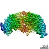
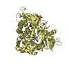
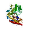
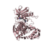
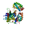
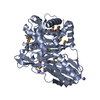
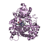
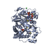
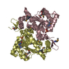
| Title | Structure of unphosphorylated Pediculus humanus (Ph) PINK1 D334A mutant | |||||||||
 Components Components | Serine/threonine-protein kinase PINK1 | |||||||||
 Keywords Keywords | TRANSFERASE / PINK1 / Kinase / Mitophagy / Parkinson's Disease / Ubiquitin / Phosphorylation / Phospho-ubiquitin | |||||||||
| Function / homology |  Function and homology information Function and homology informationautophagy of mitochondrion / positive regulation of mitochondrial fission / regulation of apoptotic process / protein kinase activity / mitochondrial outer membrane / non-specific serine/threonine protein kinase / mitochondrial inner membrane / protein serine/threonine kinase activity / ATP binding / metal ion binding / cytosol Similarity search - Function | |||||||||
| Biological species |  Pediculus humanus corporis (human body louse) Pediculus humanus corporis (human body louse) | |||||||||
| Method |  X-RAY DIFFRACTION / SYNCHROTRON / MOLECULAR REPLACEMENT / Resolution: 3.53 Å X-RAY DIFFRACTION / SYNCHROTRON / MOLECULAR REPLACEMENT / Resolution: 3.53 Å | |||||||||
 Authors Authors | Gan, Z.Y. / Leis, A. / Dewson, G. / Glukhova, A. / Komander, D. | |||||||||
| Funding support |  United States, 2items United States, 2items
| |||||||||
 Citation Citation | Journal: Nature / Year: 2022 Title: Activation mechanism of PINK1. Authors: Zhong Yan Gan / Sylvie Callegari / Simon A Cobbold / Thomas R Cotton / Michael J Mlodzianoski / Alexander F Schubert / Niall D Geoghegan / Kelly L Rogers / Andrew Leis / Grant Dewson / Alisa ...Authors: Zhong Yan Gan / Sylvie Callegari / Simon A Cobbold / Thomas R Cotton / Michael J Mlodzianoski / Alexander F Schubert / Niall D Geoghegan / Kelly L Rogers / Andrew Leis / Grant Dewson / Alisa Glukhova / David Komander /   Abstract: Mutations in the protein kinase PINK1 lead to defects in mitophagy and cause autosomal recessive early onset Parkinson's disease. PINK1 has many unique features that enable it to phosphorylate ...Mutations in the protein kinase PINK1 lead to defects in mitophagy and cause autosomal recessive early onset Parkinson's disease. PINK1 has many unique features that enable it to phosphorylate ubiquitin and the ubiquitin-like domain of Parkin. Structural analysis of PINK1 from diverse insect species with and without ubiquitin provided snapshots of distinct structural states yet did not explain how PINK1 is activated. Here we elucidate the activation mechanism of PINK1 using crystallography and cryo-electron microscopy (cryo-EM). A crystal structure of unphosphorylated Pediculus humanus corporis (Ph; human body louse) PINK1 resolves an N-terminal helix, revealing the orientation of unphosphorylated yet active PINK1 on the mitochondria. We further provide a cryo-EM structure of a symmetric PhPINK1 dimer trapped during the process of trans-autophosphorylation, as well as a cryo-EM structure of phosphorylated PhPINK1 undergoing a conformational change to an active ubiquitin kinase state. Structures and phosphorylation studies further identify a role for regulatory PINK1 oxidation. Together, our research delineates the complete activation mechanism of PINK1, illuminates how PINK1 interacts with the mitochondrial outer membrane and reveals how PINK1 activity may be modulated by mitochondrial reactive oxygen species. | |||||||||
| History |
|
- Structure visualization
| Structure viewer | Molecule: MolmilJmol/JSmol |
|---|
- Downloads & links
-Download
| PDBx/mmCIF format | 7t3x.cif.gz | 110.6 KB | Display | PDBx/mmCIF format |
|---|---|---|---|---|
| PDB format | pdb7t3x.ent.gz | 66.9 KB | Display | PDB format |
| PDBx/mmJSON format | 7t3x.json.gz | Tree view | PDBx/mmJSON format | |
| Others |  Other downloads Other downloads |
-Validation report
| Arichive directory | https://data.pdbj.org/pub/pdb/validation_reports/t3/7t3xftp://data.pdbj.org/pub/pdb/validation_reports/t3/7t3x | HTTPS FTP |
|---|
-Related structure data
| Related structure data |  7t4kC  7t4lC  7t4mC  7t4nC  6eqiS S: Starting model for refinement C: citing same article ( |
|---|---|
| Similar structure data |


-Links
 PDBj
PDBj
- Assembly
| Deposited unit | 
| ||||||||||||
|---|---|---|---|---|---|---|---|---|---|---|---|---|---|
| 1 |
| ||||||||||||
| Unit cell |
|
-Components
| #1: Protein | Mass: 52929.613 Da / Num. of mol.: 1 / Mutation: D334A Source method: isolated from a genetically manipulated source Source: (gene. exp.) Pediculus humanus corporis (human body louse)Gene: 8239562, Phum_PHUM577390 / Plasmid: pOPIN-K / Production host:  References: UniProt: E0W1I1, non-specific serine/threonine protein kinase |
|---|
-Experimental details
-Experiment
| Experiment | Method: X-RAY DIFFRACTION / Number of used crystals: 1 |
|---|
- Sample preparation
| Crystal | Density Matthews: 2.59 Å3/Da / Density % sol: 52.59 % |
|---|---|
| Crystal grow | Temperature: 281 K / Method: vapor diffusion, sitting drop / pH: 7 Details: 20% polyethylene glycol 3350, 0.2 M triammonium citrate, 0.1 M ammonium sulphate, 0.01 M magnesium chloride |
-Data collection
| Diffraction | Mean temperature: 100 K / Serial crystal experiment: N |
|---|---|
| Diffraction source | Source: SYNCHROTRON / Site: Australian Synchrotron / Beamline: MX2 / Wavelength: 0.9537 Å |
| Detector | Type: DECTRIS EIGER X 16M / Detector: PIXEL / Date: Nov 27, 2020 |
| Radiation | Protocol: SINGLE WAVELENGTH / Monochromatic (M) / Laue (L): M / Scattering type: x-ray |
| Radiation wavelength | Wavelength: 0.9537 Å / Relative weight: 1 |
| Reflection | Resolution: 3.53→44.83 Å / Num. obs: 6769 / % possible obs: 99.9 % / Redundancy: 5.7 % / Biso Wilson estimate: 80.2 Å2 / Rmerge(I) obs: 0.41 / Rpim(I) all: 0.188 / Net I/σ(I): 4.2 |
| Reflection shell | Resolution: 3.53→3.87 Å / Redundancy: 5.8 % / Rmerge(I) obs: 0.953 / Mean I/σ(I) obs: 2 / Num. unique obs: 1609 / % possible all: 99.7 |
- Processing
| Software |
| ||||||||||||||||||||||||||||
|---|---|---|---|---|---|---|---|---|---|---|---|---|---|---|---|---|---|---|---|---|---|---|---|---|---|---|---|---|---|
| Refinement | Method to determine structure: MOLECULAR REPLACEMENT Starting model: 6EQI Resolution: 3.53→44.83 Å / SU ML: 0.3182 / Cross valid method: FREE R-VALUE / σ(F): 1.34 / Phase error: 26.8819 Stereochemistry target values: GeoStd + Monomer Library + CDL v1.2
| ||||||||||||||||||||||||||||
| Solvent computation | Shrinkage radii: 0.9 Å / VDW probe radii: 1.11 Å / Solvent model: FLAT BULK SOLVENT MODEL | ||||||||||||||||||||||||||||
| Displacement parameters | Biso mean: 58.17 Å2 | ||||||||||||||||||||||||||||
| Refinement step | Cycle: LAST / Resolution: 3.53→44.83 Å
| ||||||||||||||||||||||||||||
| Refine LS restraints |
| ||||||||||||||||||||||||||||
| LS refinement shell |
|
