 Movie
Movie Controller


+ Open data

- Basic information
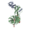
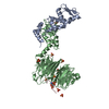
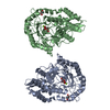
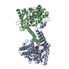
| Entry | Database: PDB / ID: 7t1z | ||||||
|---|---|---|---|---|---|---|---|
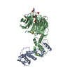
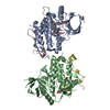
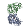
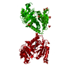
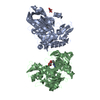
| Title | Structure of the Fbw7-Skp1-MycNdegron complex | ||||||
 Components Components |
| ||||||
 Keywords Keywords | LIGASE / Ubiquitin ligase / WD40 | ||||||
| Function / homology |  Function and homology information Function and homology informationnegative regulation of SREBP signaling pathway / negative regulation of triglyceride biosynthetic process / negative regulation of hepatocyte proliferation / regulation of lipid storage / ubiquitin recycling / ubiquitin-protein transferase activator activity / Parkin-FBXW7-Cul1 ubiquitin ligase complex / F-box domain binding / regulation of mitophagy / regulation of cell cycle G1/S phase transition ...negative regulation of SREBP signaling pathway / negative regulation of triglyceride biosynthetic process / negative regulation of hepatocyte proliferation / regulation of lipid storage / ubiquitin recycling / ubiquitin-protein transferase activator activity / Parkin-FBXW7-Cul1 ubiquitin ligase complex / F-box domain binding / regulation of mitophagy / regulation of cell cycle G1/S phase transition / negative regulation of osteoclast development / PcG protein complex / positive regulation of oxidative stress-induced neuron intrinsic apoptotic signaling pathway / phosphothreonine residue binding / positive regulation of ubiquitin protein ligase activity / Cul7-RING ubiquitin ligase complex / maintenance of protein location in nucleus / positive regulation of proteasomal protein catabolic process / Loss of Function of FBXW7 in Cancer and NOTCH1 Signaling / positive regulation of ubiquitin-dependent protein catabolic process / vasculature development / : / sister chromatid cohesion / SCF ubiquitin ligase complex / SCF-dependent proteasomal ubiquitin-dependent protein catabolic process / Prolactin receptor signaling / Association of TriC/CCT with target proteins during biosynthesis / ubiquitin ligase complex scaffold activity / lipid homeostasis / cullin family protein binding / negative regulation of Notch signaling pathway / protein monoubiquitination / positive regulation of epidermal growth factor receptor signaling pathway / ubiquitin-like ligase-substrate adaptor activity / protein K48-linked ubiquitination / : / Nuclear events stimulated by ALK signaling in cancer / molecular function activator activity / cyclin binding / ubiquitin binding / Regulation of BACH1 activity / positive regulation of protein ubiquitination / MAP3K8 (TPL2)-dependent MAPK1/3 activation / SCF-beta-TrCP mediated degradation of Emi1 / NIK-->noncanonical NF-kB signaling / Vpu mediated degradation of CD4 / Dectin-1 mediated noncanonical NF-kB signaling / Activation of NF-kappaB in B cells / Degradation of GLI1 by the proteasome / Iron uptake and transport / GSK3B and BTRC:CUL1-mediated-degradation of NFE2L2 / Negative regulation of NOTCH4 signaling / FBXL7 down-regulates AURKA during mitotic entry and in early mitosis / Degradation of GLI2 by the proteasome / GLI3 is processed to GLI3R by the proteasome / beta-catenin binding / regulation of circadian rhythm / Degradation of beta-catenin by the destruction complex / NOTCH1 Intracellular Domain Regulates Transcription / CLEC7A (Dectin-1) signaling / Constitutive Signaling by NOTCH1 PEST Domain Mutants / Constitutive Signaling by NOTCH1 HD+PEST Domain Mutants / SCF(Skp2)-mediated degradation of p27/p21 / FCERI mediated NF-kB activation / Interleukin-1 signaling / protein polyubiquitination / Orc1 removal from chromatin / Regulation of RUNX2 expression and activity / Cyclin D associated events in G1 / cellular response to UV / Regulation of PLK1 Activity at G2/M Transition / Downstream TCR signaling / rhythmic process / Antigen processing: Ubiquitination & Proteasome degradation / regulation of protein localization / chromosome / Neddylation / protein-macromolecule adaptor activity / proteasome-mediated ubiquitin-dependent protein catabolic process / positive regulation of ERK1 and ERK2 cascade / protein stabilization / protein ubiquitination / chromatin remodeling / protein domain specific binding / negative regulation of gene expression / DNA repair / DNA damage response / ubiquitin protein ligase binding / centrosome / nucleolus / protein-containing complex / nucleoplasm / identical protein binding / nucleus / cytoplasm / cytosol Similarity search - Function | ||||||
| Biological species |  Homo sapiens (human) Homo sapiens (human) | ||||||
| Method |  X-RAY DIFFRACTION / SYNCHROTRON / MOLECULAR REPLACEMENT / Resolution: 2.77 Å X-RAY DIFFRACTION / SYNCHROTRON / MOLECULAR REPLACEMENT / Resolution: 2.77 Å | ||||||
 Authors Authors | Wang, B. / Rusnac, D.V. / Clurman, B.E. / Zheng, N. | ||||||
| Funding support |  United States, 1items United States, 1items
| ||||||
 Citation Citation | Journal: Sci Adv / Year: 2022 Title: Two diphosphorylated degrons control c-Myc degradation by the Fbw7 tumor suppressor. Authors: Welcker, M. / Wang, B. / Rusnac, D.V. / Hussaini, Y. / Swanger, J. / Zheng, N. / Clurman, B.E. | ||||||
| History |
|
- Structure visualization
| Structure viewer | Molecule: MolmilJmol/JSmol |
|---|
- Downloads & links
-Download
| PDBx/mmCIF format | 7t1z.cif.gz | 143.3 KB | Display | PDBx/mmCIF format |
|---|---|---|---|---|
| PDB format | pdb7t1z.ent.gz | 100.7 KB | Display | PDB format |
| PDBx/mmJSON format | 7t1z.json.gz | Tree view | PDBx/mmJSON format | |
| Others |  Other downloads Other downloads |
-Validation report
| Arichive directory | https://data.pdbj.org/pub/pdb/validation_reports/t1/7t1zftp://data.pdbj.org/pub/pdb/validation_reports/t1/7t1z | HTTPS FTP |
|---|
-Related structure data
| Related structure data | 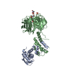 7t1yC 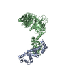 2ovpS C: citing same article ( S: Starting model for refinement |
|---|---|
| Similar structure data |

-Links
 PDBj
PDBj


- Assembly
| Deposited unit | 
| ||||||||||||
|---|---|---|---|---|---|---|---|---|---|---|---|---|---|
| 1 |
| ||||||||||||
| Unit cell |
|
-Components
| #1: Protein | Mass: 16771.787 Da / Num. of mol.: 1 Source method: isolated from a genetically manipulated source Source: (gene. exp.) Homo sapiens (human) / Gene: SKP1, EMC19, OCP2, SKP1A, TCEB1L / Production host:  | ||||||
|---|---|---|---|---|---|---|---|
| #2: Protein | Mass: 51584.789 Da / Num. of mol.: 1 Source method: isolated from a genetically manipulated source Source: (gene. exp.) Homo sapiens (human) / Gene: FBXW7, FBW7, FBX30, SEL10 / Production host: | ||||||
| #3: Protein/peptide | Mass: 2573.770 Da / Num. of mol.: 1 / Source method: obtained synthetically / Source: (synth.) Homo sapiens (human) | ||||||
| #4: Chemical | ChemComp-SO4 /   Mass: 96.063 Da / Num. of mol.: 5 / Source method: obtained synthetically / Formula: SO4 Mass: 96.063 Da / Num. of mol.: 5 / Source method: obtained synthetically / Formula: SO4#5: Water | ChemComp-HOH / |  Mass: 18.015 Da / Num. of mol.: 99 / Source method: isolated from a natural source / Formula: H2O Mass: 18.015 Da / Num. of mol.: 99 / Source method: isolated from a natural source / Formula: H2OHas ligand of interest | Y | Has protein modification | Y | |
-Experimental details
-Experiment
| Experiment | Method: X-RAY DIFFRACTION / Number of used crystals: 1 |
|---|
- Sample preparation
| Crystal | Density Matthews: 5.54 Å3/Da / Density % sol: 77.8 % |
|---|---|
| Crystal grow | Temperature: 298.15 K / Method: vapor diffusion, hanging drop / pH: 6.5 / Details: 0.1 M Bicine pH6.5, 1.6 M Li2SO4 |
-Data collection
| Diffraction | Mean temperature: 123 K / Serial crystal experiment: N |
|---|---|
| Diffraction source | Source: SYNCHROTRON / Site: ALS / Beamline: 8.2.2 / Wavelength: 1 Å |
| Detector | Type: ADSC QUANTUM 315r / Detector: CCD / Date: Apr 29, 2021 |
| Radiation | Protocol: SINGLE WAVELENGTH / Monochromatic (M) / Laue (L): M / Scattering type: x-ray |
| Radiation wavelength | Wavelength: 1 Å / Relative weight: 1 |
| Reflection | Resolution: 2.77→50 Å / Num. obs: 37654 / % possible obs: 100 % / Redundancy: 9.7 % / Biso Wilson estimate: 52.88 Å2 / CC1/2: 0.992 / Net I/σ(I): 12 |
| Reflection shell | Resolution: 2.77→2.82 Å / Num. unique obs: 1861 / CC1/2: 0.602 |
- Processing
| Software |
| |||||||||||||||||||||||||||||||||||||||||||||||||||||||||||||||||||||||||||||||||||||||||||||||||||||||||
|---|---|---|---|---|---|---|---|---|---|---|---|---|---|---|---|---|---|---|---|---|---|---|---|---|---|---|---|---|---|---|---|---|---|---|---|---|---|---|---|---|---|---|---|---|---|---|---|---|---|---|---|---|---|---|---|---|---|---|---|---|---|---|---|---|---|---|---|---|---|---|---|---|---|---|---|---|---|---|---|---|---|---|---|---|---|---|---|---|---|---|---|---|---|---|---|---|---|---|---|---|---|---|---|---|---|---|
| Refinement | Method to determine structure: MOLECULAR REPLACEMENT Starting model: 2ovp Resolution: 2.77→49.96 Å / SU ML: 0.4146 / Cross valid method: FREE R-VALUE / σ(F): 1.35 / Phase error: 24.1643 Stereochemistry target values: GeoStd + Monomer Library + CDL v1.2
| |||||||||||||||||||||||||||||||||||||||||||||||||||||||||||||||||||||||||||||||||||||||||||||||||||||||||
| Solvent computation | Shrinkage radii: 0.9 Å / VDW probe radii: 1.11 Å / Solvent model: FLAT BULK SOLVENT MODEL | |||||||||||||||||||||||||||||||||||||||||||||||||||||||||||||||||||||||||||||||||||||||||||||||||||||||||
| Displacement parameters | Biso mean: 55.55 Å2 | |||||||||||||||||||||||||||||||||||||||||||||||||||||||||||||||||||||||||||||||||||||||||||||||||||||||||
| Refinement step | Cycle: LAST / Resolution: 2.77→49.96 Å
| |||||||||||||||||||||||||||||||||||||||||||||||||||||||||||||||||||||||||||||||||||||||||||||||||||||||||
| Refine LS restraints |
| |||||||||||||||||||||||||||||||||||||||||||||||||||||||||||||||||||||||||||||||||||||||||||||||||||||||||
| LS refinement shell |
|
